


 View fulltext
Advanced Photonics
Co-Editors-in-Chief
Xiao-Cong (Larry) Yuan, Anatoly Zayats

2025
Volume: 7 Issue 5
11 Article(s)

Chao Zuo, and Qian Chen
The commentary discusses a recent report of metasurface-enhanced infrared photothermal (MEIP) microscopy, which simultaneously overcomes long-standing sensitivity and spatial resolution barriers in midinfrared spectroscopy, advanc
Aug. 29, 2025Vol. 7 Issue 5 050501 (2025)
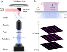
Hongyou Zhao... and Ying Gu|Show fewer author(s)
In the past two decades, optogenetic technology has developed to be the most accurate method for investigating or treating neural correlated diseases. Currently, the applications of optogenetic technology have been expanded from t
Aug. 21, 2025Vol. 7 Issue 5 054001 (2025)
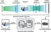
Fei Wang... and Guohai Situ|Show fewer author(s)
Computational imaging (CI) leverages the joint optimization of optical system design and reconstruction algorithms, enabling superior performance in terms of dimensionality, resolution, efficiency, and hardware complexity. It has
Sep. 03, 2025Vol. 7 Issue 5 054002 (2025)
Boris Chichkov
Quantum theory of photons based on the first quantization technique, similar to that used by Schrödinger in the formulation of quantum mechanics, is considered. First, scalar quantum mechanics of photons operating with the photon
Aug. 18, 2025Vol. 7 Issue 5 055001 (2025)

Danchen Jia... and Ji-Xin Cheng|Show fewer author(s)
Infrared spectroscopy with its rich vibrational information plays a crucial role in biochemical sensing. Metasurface-enhanced infrared spectroscopy amplifies the detection sensitivity by enhancing local electromagnetic fields. How
Jul. 15, 2025Vol. 7 Issue 5 056001 (2025)

Xuan Li... and Xiaoshun Jiang|Show fewer author(s)
Visible optical frequency combs are essential to optical atomic clocks, astronomical spectrograph calibration, and biological imaging. However, due to the limitations of the dispersion properties of available materials and the Q-f
Jul. 17, 2025Vol. 7 Issue 5 056002 (2025)

Shunshun Yang... and Linglong Zhang|Show fewer author(s)
Quantum-confined Stark effects (QCSEs), where external or built-in electric fields modify optical transition energies, have garnered significant interest due to their potential for tuning emission energies to couple with quantum d
Aug. 05, 2025Vol. 7 Issue 5 056003 (2025)
Tong Liu... and Lei Zhou|Show fewer author(s)
Holography plays a crucial role in optics, yet traditional methods require complex setups and bulky devices, being unfavourable for optical integration. Although metasurface-based holograms can be ultra-compact, holographic images
Aug. 19, 2025Vol. 7 Issue 5 056004 (2025)
Min Huang... and Hongsheng Chen|Show fewer author(s)
Robust three-dimensional (3D) recognition across different viewing angles is crucial for dynamic applications such as autonomous navigation and augmented reality; however, the application of the technology remains challenging owin
Aug. 20, 2025Vol. 7 Issue 5 056005 (2025)
Xinyang Wang... and Haiping He|Show fewer author(s)
All-inorganic CsPbBr3 perovskite polycrystalline films, renowned for their remarkable optoelectronic properties, solution processability, and enhanced stability over organic–inorganic counterparts, are emerging as next-generation
Aug. 19, 2025Vol. 7 Issue 5 056006 (2025)
Hangbo Yang... and Volker J. Sorger|Show fewer author(s)
Convolutional operations are computationally intensive in artificial intelligence (AI) services, and their overhead in electronic hardware limits machine learning scaling. Here, we introduce a photonic joint transform correlator (
Sep. 08, 2025Vol. 7 Issue 5 056007 (2025)
© Copyright 2018-2021 | Chinese Laser Press.
All Rights Reserved 沪ICP备15018463号-20