


Please enter the answer below before you can view the full text.
8-8=

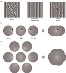
Conventional optical microscopes can achieve super-resolution and optical sectioning capabilities by replacing the source of uniform illumination with structured illumination module. Because of the configuration compatibility with the conventional wide-field optical microscope, the structured illumination microscopy (SIM) inherits the merits of non-invasiveness, low phototoxicity, low photo-bleaching, and fast imaging speed. The high spatiotemporal resolution and three-dimensional optical sectioning abilities of SIM are highly suitable for the observation of living cells or tissues. Thus, SIM has attracted continuous attention by the biomedical and optical communities. The core techniques of SIM are fast fringe generation with high contrast and high frequency, as well as fast phase shifting and fringe rotation. The digital micromirror device (DMD) based SIM (DMD-SIM) has undergone rapid development in recent years. DMD-SIM, taking the advantages of high refreshing rate, high photon flux efficiency, and insensitive to polarization, has overcome the drawbacks of the low modulation speeds of traditional devices, e.g., physical gratings and liquid crystal spatial light modulators. First, the basic principles of SIM for super-resolution and optical sectioning are introduced. Then it focuses on the DMD-SIM for generation of structured illuminations by using either beam projection or beam interference methods. Furthermore, the advances in DMD-SIM technology are reviewed, and the advantages and disadvantages of DMD-SIM are summarized. Finally, the challenges and the outlook for DMD-SIM are anticipated.
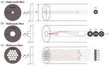
Lensless ultrathin fiber-optic endomicroscopic imaging technology based on multimode fiber or multicore fiber has made great progress in recent years, and it is expected to produce the next-generation ultra-minimal high-resolution endoscope. By spatiotemporally controlling the coherent incident light field, the influence of mode dispersion in multimode fibers or phase distortion in multicore fibers can be overcome. High-resolution focusing, imaging, and related applications can be achieved through optical fibers without using lens or scanning device at the distal end. In addition, in scenarios of lensless fiber-optic endoscopic imaging or image transmission, object information reconstruction can be achieved from the output measurements by building a physical or deep learning model. This article reviews the development of coherent optical fiber lensless imaging technology. First, it depicts the basic working principles of lensless optical fiber imaging and the imaging methods from the two perspectives of active wavefront control and passive object reconstruction. Then it introduces some advanced optical fiber imaging modalities and technologies and lists fiber imaging related applications, with discussions of general challenges encountered in this field. Finally, a brief summary is made with envision of the development roadmaps.
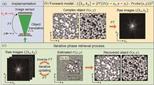
Ptychography is a coherent diffraction imaging technique and has rapidly developed over the last decade, becoming an indispensable imaging tool in most X-ray synchrotrons and national laboratories globally. In the visible light regime, optical ptychography can be divided into two: lens-based Fourier ptychography and lensless-based coded ptychography (CP). CP is a novel lensless on-chip microscopy imaging technique, and its advantages include a large field-of-view, high-resolution, aberration-free, label-free, field portability, and slow-varying phase imaging. In this review, we discuss the basic principles of CP and summarize its recent progress. Additionally, we analyze its imaging performance and highlight its biomedical applications. Finally, we conclude this review article by pointing out several directions for its future development.
The diffraction limit prevents traditional optical imaging methods from observing organelle structures and interaction between the organelles. As the imaging technique with the highest resolution among the three super-resolution techniques, single-molecule localization microscopy is an important tool for research in the field of life science. Single-molecule imaging technology with a large field of view and high throughput has been widely used in the biomedical field to observe and analyze complex biological structures and functions due to its high resolution, wide imaging range, and short imaging time. In this paper, the recent research progress of high-throughput single-molecule localization microscopy with a large field of view is reviewed from four perspectives: hardware scanning-based mosaic imaging technology, large-array sCMOS large-field-of-view high-throughput imaging technology, large-depth single-molecule positioning imaging technology, and high-throughput data analysis technology. Finally, the development direction for the large-field-of-view high-throughput single-molecule localization microscopy is prospected.
Because of the high sensitivity and specificity and unparalleled ability to quantitatively analyze tissue microenvironments, fluorescence lifetime imaging microscopy (FLIM) is widely used in the field of life sciences. However, the use of FLIM in live imaging is hindered by its slow imaging speed. Recent advancements in optoelectronics and artificial intelligence technologies have ushered in a new era for in vivo FLIM. This review introduces ways to improve the imaging speed of the time-domain and frequency-domain FLIM techniques through hardware optimization and algorithmic improvements as well as via advancements in fundamental biomedical research and clinical diagnostics. Finally, we explore the future directions of the in vivo FLIM technology.
Photoacoustic microscopy (PAM) is a noninvasive imaging technique that has undergone remarkable advancements and applications in the field of life sciences, basic medical research, and medical diagnostics. It operates on the unique principles of detecting photoacoustic signals and reconstructing them to create high-resolution, in-depth structural and functional images. This paper offers a comprehensive overview of the developmental background and unique principles behind photoacoustic microscopy. As we delve deeper, we explored various methods that have been employed to boost imaging performance. These include, but are not limited to, optical enhancement, acoustic enhancement, exploitation of artificial intelligence to augment the entire process. Moreover, there has been a significant focus on the harmonious integration of optics and acoustics in PAM. Ultimately, we discussed the extensive applications of current PAM in modern biomedical research and provided insights into the future developmental trends of this technique.
Coherent Raman scattering (CRS) is an important label-free chemical imaging modality that enhances Raman scattering signals via the coherent excitation of vibrational modes in molecules. Coherent enhancement considerably increases the speed of imaging and has various applications in many fields, such as material science, biochemistry, tumor diagnosis, and pharmacokinetics. The emergence of ultrafast pulsed lasers with sub-picosecond pulse durations has introduced novel pathways for CRS through the impulsive excitation of numerous vibrational modes in a synchronous and coherent manner. After discussing the fundamental principles of CRS, this study introduces the main methods of time-domain CRS. Moreover, the latest advances and applications of time-domain stimulated Raman scattering (SRS) and coherent anti-Stokes Raman scattering (CARS) have been discussed.
Stochastic optical reconstruction microscopy (STORM), a super-resolution imaging technology based on immunofluorescence, has gained popularity owing to its straightforward principle, simple optical path, and excellent spatial resolution. However, the enhanced resolution demands greater specificity from antibodies. While direct labeling with primary antibodies is an option, indirect labeling using a"primary + secondary antibody"combination is more commonly employed in practical applications. Considering the issue of species specificity with secondary antibodies, preadsorption is required to increase specificity during production. In this study, we aim to explore the effect of secondary antibody species specificity on dual-color STORM imaging. In the classical erythrocyte skeleton model, based on the mutually exclusive localization between the N-terminal and C-terminal of β-spectrin, we performed two-color STORM imaging of them by labeling them with low- and high-adsorption secondary antibodies, respectively. A comparison with cross-correlation data from simulations revealed that low-adsorption secondary antibodies led to colocalization artifacts in the STORM results. Furthermore, when employing high- and low-adsorption secondary antibodies, two-color STORM results of CD47 and PD-L1 on the MDA-MB-231 cell membrane indicated no colocalization relation between the two proteins. Therefore, this study provides an innovative super-resolution imaging strategy for evaluating secondary antibody species specificity using an erythrocyte skeleton structure model. Additionally, this approach paves the way for accurate identification of biomolecular interactions through dual-color STORM imaging.
Raman spectroscopy has the advantages of being noninvasive, fingerprint free, water background free, simple sampling preparation, and high spectral resolution; therefore, it has become a popular analytical technique in many research fields. In particular, the Raman spectroscopy can intrinsically capture biochemical information related to normal physiology or abnormal pathology in biomedicine. Additionally, various biological and clinical samples, such as cells, tissues, body fluids, and microbiology can be easily studied using the Raman spectroscopy without complex sample preparation. Thus far, the Raman spectroscopy has shown its high potential for biomedical applications. This review mainly includes an introduction to the basic mechanisms of spontaneous Raman and surface-enhanced Raman scatterings, followed by a description of the analysis procedures and algorithms for obtaining Raman data (spectral and imaging). Further, a brief summary of the applications of noncoherent Raman microscopy in biomedicine in recent years is presented, and its current challenges and future directions are discussed.
Raman microscopic imaging has emerged as an important research tool in life sciences because it does not require sample preparation and is non-destructive, non-invasive, and insensitive to aqueous solutions. It enables the characterization of biochemical components and sample distributions at a micrometer or nanometer scale. As the research on complex biological samples increases, Raman microscopic imaging holds potential for the dynamic stereoscopic observation of molecular composition and distribution in biological samples. This paper systematically examined recent advancements in 3D Raman microscopic imaging, including technical approaches, improvement strategies, and experimental results of various 3D imaging methods based on spontaneous Raman scattering, coherent Raman scattering, surface-enhanced Raman scattering, and Raman tags. Further, the progress of the applications of different imaging techniques in cell biology and developmental biology was summarized. Finally, the challenges and development prospects of different 3D Raman microscopic imaging techniques in the application of biomedical optical microscopic imaging technology were outlined.
Organisms are composed of molecules typically arranged and oriented in order. The structures of organisms are related to the positional distribution of molecules and their spatial orientation. Polarized fluorescence microscopy (PFM) is such an imaging tool that facilitates the sense and observation of the molecule orientation in biological samples by utilizing fluorescence polarization, enabling the revealing of functional and metabolic information beyond fluorescence intensity provided by more traditional fluorescence microscopy. In this review, we first briefly describe the theory and principle of PFM and then introduce several implementations of PFM on different microscopic modalities. We later summarize its applications in the field of biomedical research and finally discuss its potential future directions.
Optical thin films are widely used in optical instruments and measurement and control technology, thereby affecting various aspects of our life and making it diverse and colorful. Unlike conventional applications of optical thin films, this review focuses on the combination of optical thin films with optical microscopy imaging techniques. The main research plan is to develop planar thin film photonic devices for unmarked microscopic detection based on noble metal thin films loaded with surface plasmon waves and dielectric multilayer thin films with photonic bandgap structures. Owing to its planar structure and mature manufacturing process, this type of photonic thin film element can be made compatible with conventional bright field and wide field microscopic imaging systems, which makes it an ideal substrate for a test sample or can be used as a plugin for the imaging system. Such devices can utilize the near-far field interaction characteristics of thin films and light waves to regulate the illumination field of the system for achieving dark field illumination, total internal reflection illumination, and edge enhanced illumination. The contrast and detection sensitivity of imaging can be improved by changing the lighting method, leading to the development of multimodal, unmarked optical microscopy imaging and sensing systems with high sensitivity and contrast. To utilize the characteristics of simple structure, wide field, high sensitivity, and unlabeled imaging of this system, it has been applied to the field of environmental photonics for in situ, real-time, and nondestructive investigation of the hygroscopic growth process of a single ultrafine particle in a real atmospheric environment. Thus, this imaging system is expected to provide strong scientific support and technical tools for tracing of and further research on atmospheric haze.
Quantitative evaluation has emerged as a critical aspect of industrial digital image quality assessment, particularly in the field of optical microscopy. It primarily assesses the quality of optical microscopic images by analyzing their features and attributes. In recent years, the evolution of various optical microscopic technologies has highlighted the importance of quantitative image evaluation. Furthermore, it plays a pivotal role in guiding the overall image processing workflow. This review comprehensively examines quantitative evaluation indicators and their associated algorithms, focusing on their model structures and performances. Moreover, it addresses the current challenges in this domain, outlines the latest developments in the quantitative assessment of optical microscopic images, and suggests future research directions.
Light sheet microscope is an important tool for three-dimensional imaging owing to its powerful optical section capability, fast imaging speed, and low phototoxicity. This microscope typically uses two objectives that are placed perpendicular to each other for illumination and imaging; however, this structure imposes spatial constraints and prohibits the use of an imaging objective with high numerical aperture. These limitations were resolved using single-objective light sheet microscopy techniques, such as oblique plane illumination and micromirror microdevice reflection, demonstrating potential for high-resolution and volumetrically high-speed imaging. Furthermore, these techniques can be combined with various other techniques, such as super-resolution microscopy, which has made great progress in recent years. This review article introduces the principles, key performance enhancements, and biomedical applications of single-objective light sheet microscopy.
Super-resolution microscopy with nanoscale spatial resolution has become an important imaging tool in life science research. As a super-resolution technique, single-molecule localization microscopy enables us to localize, identify, and study the unique behaviors of single molecules. At the single-molecule level, the emitted fluorescence signal is highly anisotropic. Resolving the polarization or three-dimensional orientation of single fluorescent molecules is an emerging field in super-resolution microscopy. In this review, we describe the three-dimensional orientation of single molecules through super-resolution imaging techniques. These techniques include fluorescence polarization microscopy and single-molecule orientation imaging through point spread function engineering. Furthermore, we discuss other polarization super-resolution imaging approaches for the applications of live cells and single nanoparticle studies. Finally, we discuss the potential challenges and future research needs of single-molecule orientation localization microscopy. These challenges and requirements can provide in-depth insights into future research in life sciences.
The last two decades have witnessed the invention and development of super-resolution microscopy (SRM) that breaks the diffraction limit of light and pushes the fluorescence microscopy resolution to several nanometers. While SRM is widely used in biological studies, such as resolving subdiffraction structures, molecular mapping, tracking single molecules, probing protein-protein interactions, and observing organelle dynamics, its direct application in translational medicine, such as disease diagnosis, is still preliminary. Despite their small size, cilia play a crucial role as organelles in cell signaling and motility, with defects in cilia leading to ciliopathy. Similar to other miniature organelles and macromolecular complexes, cilia are ideal for super-resolution imaging. In this review, we will 1) introduce cilia and ciliopathy, 2) show how SRM extends our knowledge of cilia, and 3) focus on how SRM improves the diagnosis of motile ciliopathies.
Photoacoustic imaging (PAI), a biomedical imaging mode that combines the high contrast of optical imaging with the deep penetration of ultrasonic imaging, has developed rapidly in recent years. Among them, photoacoustic microscopy (PAM), as one of the important implementation methods of photoacoustic imaging, can achieve micron-level or even hundreds of nanometer-level resolution on the millimeter imaging depth, and can achieve high-resolution imaging of biological tissue structure, function, and molecules, and has been widely used in clinical diagnosis, skin disease detection, ophthalmology, and other fields. In this paper, the working principle and implementation of PAM are first introduced, and then the research progress of portable PAM technology is reviewed from the handheld and semi-handheld, brain wearable, and integrated multi-mode. Then, the challenges faced by the portable PAM technology are discussed, and finally the prospects are summarized.
Optical super-resolution imaging technology, acknowledged by the Nobel Prize in Chemistry for transcending optical diffraction limits, has revolutionised life science research with its groundbreaking observation scale. However, conventional super-resolution fluorescence microscopes face challenges, requiring intricate optical systems that often result in significant phototoxicity and low temporal resolution, limiting their widespread use in biomedical research. Therefore, research teams actively seek alternative fluorescent probes with near-infrared capabilities, high brightness, and resistance to photobleaching, aiming to extend the application of super-resolution microscopy in biomedical research. Rare-earth nanomaterials, renowned for their exceptional physicochemical properties such as anti-Stokes spectral shift, lack of background noise, resistance to photobleaching, photostability, low toxicity, and high imaging penetration, have emerged as stable and superior inorganic fluorescent probes. This review paper provides a brief overview of the luminescent mechanism of upconversion nanoparticles, exploring the primary constraints in achieving photon upconversion in nanostructured materials. Additionally, it highlights the applications and advantages of lanthanide-doped upconversion nanoparticles in super-resolution biological imaging, molecular detection, and other domains. These advantages encompass reducing laser power requirements, addressing technical challenges in coupling, improving laser scanning imaging resolution and speed, and enhancing multiplexing imaging efficiency. This paper concludes by emphasizing significant challenges in particle synthesis, proposing feasible improvement measures, and outlining prospects for future development. It establishes a robust theoretical foundation and provides technical support for the widespread integration of rare-earth nanomaterials in the field of life imaging sciences.
In recent decades, the advent of light sheet fluorescence microscopy as an innovative technique in fluorescence microscopy has significantly enhanced the high spatiotemporal resolution imaging capabilities of tissue and cellular structures and functions in life science research. Compared to traditional epi-fluorescence microscopy techniques, light sheet microscopy illuminates biological samples selectively, greatly improving photon utilization efficiency, reducing phototoxicity, and significantly increasing imaging speed. Since its introduction, light sheet microscopy has gradually expanded its application field in life science research, ranging from embryology and neuroscience to tumor studies, among others. It can not only be used to observe the basic structures of cells and tissues but also for real-time monitoring of dynamic changes in biological processes. Furthermore, its multiscale characteristics make it suitable for observations across multiple scales from macro to micro. This article reviews the applications and developments of light sheet microscopy in high-throughput imaging, high-precision imaging, and usability, aiming to provide life science researchers with comprehensive understanding and reference, and to promote its application and development in more fields.
To investigate the feasibility of label-free imaging using flat-field quantitative phase microscopy for observing mitochondrial dynamics in mesenchymal stem cells (MSCs) derived from bone marrow, SD rat bone marrow MSCs were isolated and cultured. Following passages and purification, they were seeded into confocal culture dishes and placed under a flat-field quantitative phase microscope developed by our team. Label-free observations were conducted over an extended period after confirming mitochondrial characteristics using fluorescence and phase dual channels. The mitochondrial division, fusion processes, and mitochondrial changes during cell apoptosis observed using the flat-field quantitative phase microscope were analyzed. The mitochondria observed via unlabeled flat-field quantitative phase microscopy exhibited complete overlap with fluorescently labeled mitochondria, demonstrating the microscope's capability to visualize mitochondria and conduct unlabeled high-resolution imaging. Moreover, the flat-field quantitative phase microscope facilitated long-term unlabeled observations of bone marrow MSCs under cultivation conditions, enabling high-resolution recording of mitochondrial division and fusion processes. Furthermore, we used the flat-field quantitative phase microscope to record mitochondria changes under CCCP treatment for the first time, visually presenting the apoptosis via mitochondrial pathway. The flat-field quantitative phase microscope allows for prolonged, unlabeled, high-resolution observations of cultured cells, offering a new tool for investigating mitochondrial dynamics.
An automatic exposure optimization method for mitigating overexposure, slow convergence speed, and instability in HDMI digital microscopy cameras due to the inconsistent reflection levels of different targets during observation is proposed. Compared with a traditional automatic exposure control method, the proposed method facilitates exposure control based on the region of interest, metering, and brightness statistics on any area in RAW images, thus expanding the dynamic range of brightness statistics and facilitating exposure control for different targets. In addition, the method improves the accuracy of brightness statistics and mitigates overexposure caused by extremely bright or dark targets. A variable exposure adjustment step size is set for the dynamic adjustment of exposure step size according to the current brightness level, achieving coarse and fine adjustment of exposure to balance exposure convergence speed and exposure accuracy. Finally, experiments were performed using actual cameras. The proposed method reduced the exposure deviation by half compared with the traditional automatic exposure method. The exposure convergence time was shortened by more than half, and the fastest exposure convergence was achieved within four frames of an image.
In the process of microscopic imaging, the system's depth-of-field limitation results in considerable differences in the focused positions across various planes along the axial direction. This leads to partial overlap of focused regions among microscopic images from different planes. Current multifocus fusion algorithms often struggle to simultaneously extract and merge the sharpest focused parts from multiple microscopic images. Hence, this article proposes a multifocus microscopic image fusion algorithm. First, a Gaussian-like four-neighborhood gradient operator was constructed and combined with fast guided filtering to extract high-frequency focus information. Additionally, a small region focus measurement method was introduced to enhance the extraction for high-frequency focus information from sharply focused regions considering the overlap of focus information and the substantial number of pixels in wide-field microscopic image sequences. This method effectively fuses the best focus points from multiple images. Through experiments, three sets of microscopic multifocus image sequences covering the diagonal fields-of-view of 4 mm and 2 mm were captured and testing was conducted. Through comparative analysis against five commonly used multifocus image fusion algorithms, our algorithm yields an average improvement in the peak signal-to-noise ratio of 2.4772 and surpasses a structural similarity index of 0.9400. These results exhibit superior fusion effects obtained by the proposed algorithm in the focused regions, rendering fused images enriched in details and high clarity. This algorithm meets the accuracy requirements for multifocus image fusion in applications involving large field-of-view microscopic images.
Current detection modules in microspectral imaging systems primarily comprise push-broom spectral imagers, which cannot observe dynamic microscopic samples. In this study, leveraging the capabilities of metamaterial broad-spectrum-modulated spectroscopic imaging technology, a snapshot spectroscopic camera was developed and used as a detector module to form a novel snapshot microscopic spectroscopic imaging system with a microscope module. This system enables real-time acquisition of spectral curves and image information from samples. Additionally, we used the developed system to obtain the absorption spectral curves of different algae and further used a image segmentation recognition algorithm based on a support vector machine to recognize dynamic algae samples in water. A total of 80 samples were tested in this experiment, yielding 100% accuracy and 65.52% recall for the prediction results. Thus this result forms the foundation for the application of snapshot spectral imaging technology in the field of microscopy.
Chromatic confocal microscopy (CCM) combines the high spatial resolution of confocal microscope and the high wavelength resolution of spectral analysis. By virtue of the high precision, strong applicability, and nondestructive detection, it is widely used in the fields of industrial production, biomedicine, semiconductor chips, and other fields. This paper first introduces the principle of point chromatic confocal system and points out its drawback of low detection efficiency. Second, for improving the key performance indexes of chromatic confocal microscopy, the main achievements made in light source, dispersive objective lens, and spectral signal detection are described, and qualitative comparisons are made between various types of light sources. Subsequently, the scanning methods of chromatic confocal microscopy are demonstrated, the relevant research progress is sorted out, and the advantages and disadvantages of the relevant methods are summarized. Finally, the future developments of chromatic confocal microscopy are also prospected.