


Biomedical imaging technology can assist doctors in understanding morphology, function, metabolism changes in human bodies and serves as an indispensable tool in the diagnosis and treatment for a diverse of diseases such as cancers and strokes. Compared to x-ray based computed tomography (CT) with ionizing radiation hazards, magnetic resonance imaging (MRI) with strong magnetic fields exposure, and ultrasound imaging (USI) with limited tissue contrast, photoacoustic tomography (PAT) is a noninvasive hybrid imaging technique that combines the advantages of high optical contrast and low tissue attenuation of ultrasound. The excellent optical contrast and large penetration depth allows the acquisition of rich structural and functional information for angiography and molecular imaging in biomedical research and clinical diagnosis.
Photoacoustic computed tomography (PACT) is one of the modalities of PAT, which employs diffused light to illuminate the whole imaging area, and reconstructs two- (2-D) or three-dimensional (3-D) images of biological tissues using the ultrasound signals received at different locations. Piezoelectric (PbZrxTi1-xO3, PZT) ultrasound transducer is widely used in PAT by converting ultrasound waves into electrical signals in photoacoustic imaging. As the sensitivity of the ultrasound transducer scales down with its element size, relatively large size transducer is preferred for sufficient sensitivity but sacrifices the acceptance angle. This leads to the information loss, artifacts and resolution degradation for the PAT. While several research groups have improved the acceptance angle by adding acoustic convex lenses to the transducer, the mismatch in the acoustic impedance between the materials causes ultrasound loss and reverberation. Alternative solution is to customize negatively-focused ultrasound transducer, but the complexity of fabrication hinders the mass application. To address the above issues, the research group from Jinan University has designed a fiber-optic negativity-focused ultrasound detector and applied it for linear-scanning PACT, as shown in Fig. 1.
.png)
Fig. 1. Schematic of PACT based on the fiber-optic negatively-focused ultrasound detector for stroke model study.
Utilizing the flexibility and low bending loss of optical fibers, the optical fiber is bent into a convex shape, forming a negative focus without any additional acoustic lenses or customization. This fiber-optic negatively-focused ultrasound detector has exhibited a large acceptance angle of 120° and a low detection limit of 5.4 Pa. The fiber-detector-based linear-scanning PACT system have realized noninvasive mouse brain imaging with a depth of over 7 mm and a nearly-isotropic spatial resolution of ~ 130 μm. The system has been applied for imaging a mouse model of cerebral hemorrhage, suggesting its great potential for biomedical research and clinical diagnostic applications. Relevant research results were recently published in Photonics Research, Volume 12, Issue 12, 2024. [Hexiang Xu, Zitao Chen, Yuhan Wu, Chengtian Hou, Jun Ma, Bai-Ou Guan, "Noninvasive high-resolution deep-brain photoacoustic imaging with a negatively focused fiber-laser ultrasound transducer," Photonics Res. 12, 2996 (2024)]
The schematic diagram and photograph of the fiber-optic negatively-focused ultrasound detector are shown in Fig. 2(a). Two fiber Bragg gratings (FBGs) are inscribed in a piece of Er/Yb-doped fiber using 193-nm ultraviolet (UV) light and the phase-mask method to form an optical laser cavity. By optimizing the inscribing parameters, a fiber laser cavity with an effective length of 30 mm and a stable laser output is obtained. Highly sensitive ultrasound detection is achieved by demodulating the beat frequency variation of the output laser. A fixture is then lab-designed to bend the optical fiber at a certain angle to form a negative focus and a synthetic aperture focusing technique (SAFT) is employed to convert this focus into a virtual point detector, thus increasing the ultrasound acceptance angle as shown in Fig. 2(b).
Fig. 2(c) shows the coronal section images of the mouse brain obtained under different acceptance angles. With small acceptance angles less than 60°, only structures parallel to the scanning direction (horizontal) can be observed. As the acceptance angle increases, more vertical vascular structures, such as the rostral rhinal vein (RRV), become clear. In addition, as shown in the lower-left image of Fig. 2(d), a 125-μm-diameter optical fiber inserted in the mouse brain can be captured, showing the potential of the PACT system also for needle-guidance applications. The lower-right image of Fig. 2(d) shows the coronal section of the mouse brain with hemorrhagic thrombus. The photoacoustic signals in the left thrombus region is increased by 2-4 times as compared to the right side without cerebral hemorrhage.
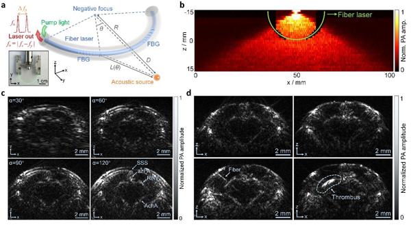
Fig. 2. (a) Schematic of the fiber-optic negatively-focused ultrasound detector. FBG, fiber Bragg grating. Inset: physical photography of the fiber-optic negatively-focused ultrasound detector. (b) The experimental results of the spatial response of the fiber-optic negatively-focused ultrasound detector. The green curve indicates the detector. (c) Reconstructed images by the PACT system for the same fiber-optic ultrasound detector with acceptance angles of 30, 60, 90, and 120 deg. SSS, superior sagittal sinus; azPA, azygos pericallosal artery; RRV, rostral rhinal vein; AchA, anterior choroidal artery. (d) Left: brain images of the mouse before and after insertion of an ink-coated optical fiber. Right: brain images of the mouse before and after the blood injection for inducing the intracerebral hemorrhage.
"Optical fiber sensors have advantages of high unit sensitivity, large bandwidth, small size, flexibility, as well as EMI resistance. The optical fiber is bent here to form a negatively focused ultrasound detector, providing both high sensitivity and a large acceptance angle for noninvasive high-resolution imaging of the mouse brain and intracerebral thrombus," explains Associate Professor Jun Ma from Jinan University, the corresponding author of the article.
The fiber-optic negatively-focused ultrasound detector currently has an acceptance angle of ~ 120°, which is mainly limited by the mechanical strength of the optical fiber and the stability of the laser output. In the future, optimizing the fiber preparation process will be carried out to further improve the acceptance angle for omnidirectional ultrasound detection and thus PAT with higher performance.