


Please enter the answer below before you can view the full text.
9-7=

The retrieval of phase information from the complex amplitude of light waves is a pivotal area of research in various scientific and engineering fields, as phase carries essential information about the propagation of light, profoundly influencing the advancement of imaging and intelligent sensing technologies. Phase recovery wavefront reconstruction techniques, leveraging advanced optimization algorithms and specialized imaging setup, extract the challenging-to-detect phase information from intensity data gathered by imaging sensors. Phase recovery wavefront reconstruction technique is one of the key methods enabling the exploration of both micro and macro worlds, extensively applied in diverse fields such as biological microscopy, industrial inspection, and astronomical observation. This paper begins by outlining both interferometric and non-interferometric wavefront reconstruction techniques and their respective applications. Subsequently, it reviews the fundamental principles and developmental trajectory of phase retrieval algorithms in wavefront reconstruction. This encompasses a preliminary exploration of prevalent phase retrieval methodologies, including alternating projection phase retrieval algorithms, ptychography imaging, and phase retrieval wavefront reconstruction techniques integrating modulation constraints and deep learning. The article concludes by summarizing the entire discourse and outlining prospective directions for the future development of phase retrieval wavefront reconstruction technology, which include the enhancement of algorithms and the development of imaging systems and devices.
Cancer poses a significant threat to human health, with tumor invasion and metastasis being the leading causes of mortality. Circulating tumor cells (CTCs) and other circulating entities are pivotal in the metastasis process. Consequently, tracking these cells and particles is critical for understanding metastasis. Optical in vivo flow cytometry (IVFC), an innovative laser-based technique, facilitates non-invasive tracking of CTCs and tumor-associated particles, proving invaluable in cancer researches, especially in metastasis studies. This study focuses on the applications of IVFC in tumor metastasis research. We discuss the detection principles of IVFC, highlighting various optical techniques such as fluorescence emission, photoacoustic effects, and computer vision. The categorization of IVFC technologies, including fluorescence in vivo flow cytometry, photoacoustic flow cytometry, and in vivo imaging flow cytometry, is detailed. We review its application in research on different cancers such as liver, prostate, breast cancer, and melanoma. The paper concludes with future prospects for IVFC in studying tumor metastasis.
Compared with two-dimensional imaging, three-dimensional (3D) LiDAR imaging has the advantages of rich information, good initiative, and strong anti-interference ability, and it has been widely used in reconnaissance, unmanned vehicle, aerospace, etc. Recently, 3D LiDAR imaging developed rapidly from two aspects. On the one hand, the manufacturing capability of APD as a typical core device has been improved, and the imaging efficiency of LiDAR has been significantly increased. On the other hand, with the continuous improvement of 3D imaging methods (scanning/non-scanning), their comprehensive performance has also been dramatically improved. This paper provides support for advancing the application of 3D imaging by summarizing the current research status of 3D LiDAR imaging, discussing the problems and proposed solutions faced in the present stage.
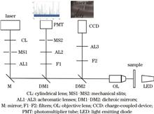
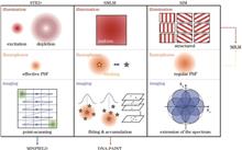
Fluorescence super-resolution microscopy has undergone several rounds of innovation and development since the 1990s. The spatial resolution of fluorescence super-resolution microscopy exceeds the diffraction limit by a wide margin, achieving a lateral resolution below to ~20 nm and enabling imaging and tracking at the molecular level. The latest generation of ultrahigh-resolution microscopy is the product of extensive development and innovative combination of conventional super-resolution techniques. This review aimed to introduce the new generation of fluorescence super-resolution microscopy at a sub-20-nm scale lateral resolution, and we also elucidated their similarities and differences compared with the conventional super-resolution techniques. Additionally, this review explored how emerging technologies optimize optical systems, scanning strategies, and sample preparation to improve resolution. Finally, we discussed the potential applications and development of high-resolution fluorescence microscopy in biomedical research.
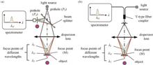
Precision measurement technology is the cornerstone of the rapid advancement of advanced manufacturing. Chromatic confocal sensors, with technological advantages such as high measurement accuracy, rapid detection speed, and high system integration, have become one of the precision measurement technologies currently garnering significant attention in the field of advanced manufacturing. This article begins by introducing the principles of chromatic confocal measurement and analyzing the crucial components comprising the chromatic confocal sensor. Subsequently, focusing on point chromatic confocal sensors, it provides an overview of the research progress in key technologies such as the dispersive objective lens, broad-spectrum light source, spectral detection device, and spectral processing algorithms that constitute the sensor. In the case of line scanning chromatic confocal sensors, it summarizes the critical technologies related to scanning methods, optical path structures, spectral detection devices, and methods for processing spectral information. Finally, it concludes by summarizing the current research focus, challenges, and future technological development directions of chromatic confocal sensors.
Holographic stereograms can be generated through optical printing and computer-generated methods, boasting uncomplicated production and authentic visual effects, which are anticipated to find applications in extensive holographic displays, augmented reality (AR), and virtual reality (VR). This paper gives an overview of the development of printing methods and the modernization of printing devices for holographic stereogram printing, as well as the iteration of computing methods and the acceleration of computational speeds in computational holographic stereograms. Finally, existing challenges are discussed and the future development of holographic stereograms is outlined.
To overcome the disadvantages of slow measurement and computation in scanning computational imaging systems, this paper summarizes several rapid computational imaging techniques and introduces methods for enhancing measurement and computation speeds. It delves into computational optical imaging methods based on light field modulation, highlighting various approaches such as axial scanning, transverse scanning, multiwavelength scanning, scattering media, and multidistance techniques. Furthermore, it explores fast quantitative phase imaging techniques, including the standard quantitative phase imaging method, rapid variant based on the Kramers-Kronig relation, computational imaging method using diagonal spread sampling, and single-frame computational imaging method employing symmetrical illumination. Additionally, it covers autofocus technologies, detailing the classification of autofocus technology, its core algorithms, the autofocus method based on the Tanimoto coefficient and the absolute value of the polyphase gradient, and the rapid autofocus method based on feature region extraction and subdivision search.
Three-dimensional (3D) shape measurement technique using structured light has garnered widespread attentions and applications in traditional manufacturing industries due to its high precision and non-contact advantages. With the rapid development of emerging fields such as smart manufacturing and artificial intelligence, there is a growing demand for efficiently acquiring high-precision 3D data sources. Phase error compensation technique, as a crucial step in achieving high-precision measuring result using structured light, plays a pivotal role in improving the accuracy and efficiency of the measuring system. In this context, this paper provides a brief introduction to the principles of phase shifting profilometry and the various forms of phase errors caused by different error sources. Then, it categorizes and discusses such methods for compensating different types of errors, optimization directions, and suitable scenarios. Finally, the paper concludes by summarizing the challenges and potential development trends of phase error compensation technique based on phase-shifting fringe analysis.
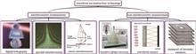
Analysis of the dynamic function of neural circuits is the focus and difficulty of current brain science research. Recently, miniaturized microscopic imaging technologies provide important research tools. Compared with two-photon fluorescence imaging and fiber photometry, miniaturized microscopic imaging systems can perform long-term, subcellular resolution, real-time imaging in freely moving animals. In the past ten years, scientists have successively developed wearable one-photon and multi-photon imaging systems with high stability. In order to improve performance and expand applications, they continuously optimized systems from the aspects of probes, optoelectronic components, data transmission and etc. In this paper, we will analyze and discuss the imaging principle, basic structure, system optimization, application scheme, and future development of miniaturized microscopic imaging systems. We summarize the research progresses in various directions, aiming to provide a reference for technology improvement and application expansion of neuroscience.
As biomedical research delves deeper into the intricacies of tissue structure and function, the demand for high-resolution, high-signal-to-noise-ratio deep-tissue imaging technologies has become critical. Traditional microscopy, limited to two-dimensional, transparent thin biological samples, falls short in satisfying the current requirements for three-dimensional deep-tissue imaging in biomedical sciences. In contrast, light-sheet fluorescence microscopy distinguishes itself through its attributes—low photodamage, rapid acquisition, extensive field of view, and volumetric imaging—establishing it as a cornerstone among biologists. Despite these advantages, the intrinsic high-scattering characteristics of biological tissues pose formidable challenges to achieving deep imaging. This review focuses on recent progress in light-sheet fluorescence microscopy for deep tissue imaging, emphasizing strategies that overcome challenges associated with high-scattering samples, so as to provide valuable insights to researchers in related fields, assisting them in developing a preliminary comprehension of the latest innovations and future possibilities inherent in this cutting-edge technology. Firstly, the basic principles of light-sheet fluorescence microscopy and the causes and effects of high-scattering absorption characteristics are explained. Subsequently, recent progress in enhancing tissue penetration depth and addressing issues such as light scattering and absorption is further examined. Finally, the development prospects and potential applications of light-sheet fluorescence microscopy imaging technology with high penetration depth and anti-scattering ability are discussed.
Quantitative phase contrast microscopy facilitates high-contrast and quantitative phase imaging of transparent samples, eliminating the need for fluorescent labeling, making it pivotal for observing dynamic processes in living cells. Traditional methods, however, require three phase-shifted interferograms to generate a quantitative phase image, resulting in time-intensive procedures. This study introduces a novel phase reconstruction approach for quantitative phase contrast microscopy, leveraging a two-channel convolutional neural network. This innovative method achieves quantitative phase image retrieval from only two phase-shifted interferograms, enhancing imaging speed by 1.5 times and reconstruction speed by an order of magnitude compared with traditional approaches. In our experimental setup, the network was trained using COS7 cell data. The trained network successfully reconstructed quantitative phase images of 3T3 cells, demonstrating its applicability for accurate and robust phase reconstruction across different cell types. This method holds promise as a powerful tool for real-time, high-resolution observation of dynamic living cells and the interaction networks of sub-cellular organelles.
Terahertz waves possess remarkable penetrability, capable of passing through a wide range of non-metallic, nonpolar materials without causing ionization. This unique characteristic makes them particularly useful in the field of computed tomography, enabling the capture of detailed external and internal structures of samples along their three-dimensional absorption coefficient distributions. In this process, the sinogram for each vertical layer is generated through raster scanning at different projection angles. These sinograms are then transformed into sectional images using filtered back projection and other algorithms based on the Fourier central slicing theorem. As the field has developed, a diverse array of illumination modes, imaging configurations, and reconstruction algorithms have been proposed. These advancements serve to accommodate the development of terahertz imaging instruments and components, as well as the expanding range of application scenarios. This study reviews the principals of terahertz computed tomography. We provide a comprehensive review of its recent research progress, focusing on enhancing three key areas: reconstruction quality, imaging resolution enhancement, and data acquisition efficiency.
Pipeline robots are vital tools for detecting and assessing pipeline damage in complex systems. Incorporating imaging systems into their forward trajectory enables these robots to navigate and observe the internal environment of pipelines. However, this positioning often results in pipeline walls information appearing at the edges of the image sensor, where lens distortions substantially impact the image quality. This distortion reduces the precision of damage detection and complicates the quantification of damage. Additionally, integrating additional imaging systems to observe the walls would substantially increase the robots payload and volume, which is a critical issue for small-scale pipeline robots. To address these challenges, we developed a miniaturized wall imaging system specifically for pipeline robots. Our approach involves careful component selection, optimization of the optical system, and integration using 3D printing, resulting in a compact system measuring 25 mm×30 mm×12 mm and offering an optimal lateral resolution of 15.63 μm. Furthermore, we constructed a miniaturized pipeline robot using this system, demonstrating its effectiveness in imaging and quantification. This innovative system can be integrated into various pipeline robots, thereby enhancing their capabilities in capturing detailed information about pipeline walls.
The paper explores the use of liquid crystal (LC) technology in XR near-eye display systems. It elaborates on how LC materials, through controlled light polarization and wavefront manipulation, contribute to the development of varifocal LC lenses. These lenses are notable for their potential to improve user experience in XR applications. The document also discusses the challenges in manufacturing LC lenses, particularly in optimizing lens thickness and performance.
High-speed optofluidic imaging is an emerging interdisciplinary technology that seamlessly integrates high-speed optical imaging and microfluidics, which is capable of realizing high-resolution, high-throughput, high-content imaging, and quantitative analysis of biological organisms in high-speed and complex fluid environments. High-speed optofluidic imaging has exhibited promising application potential in bioenergy, food science, drug screening, disease diagnosis, and many other fields. In this review, we introduce the basic principles, key techniques, and recent advances of high-speed optofluidic imaging. The future prospects and challenges of high-speed optofluidic imaging are also discussed.
Solar-induced chlorophyll fluorescence of vegetation is an important parameter in evaluating plant photosynthesis. A hyper-spectral imaging spectrometer prototype is presented for the fluorescence observation of vegetation in a wide area. The main transmission optical system of the imaging spectrometer is based on the key dispersive element composed of prism and volume phase holographic transmission grating. This form could realize high performances under the high numerical aperture (0.25). The prototype has an angle spatial resolution of 1 mrad, a spectral resolution of 0.3 nm, and an signal-to-noise ratio better than 100 in the waveband of 670‒780 nm (it can be extended to 650‒800 nm) and the full field of view of 20°. The results of the design, test results of the prototype and analysis results of application data show that the imaging spectrometer can satisfy the requests of the design. The instrument can supply important scientific data for the agriculture monitoring and forests and carbon cycle observation, and it can be considered as a new observation method for the land vegetation photosynthesis.
In a scattering environment, the optical imaging technology often encounters considerable degradation in image quality, exhibiting diminished contrast for details. This limitation has hindered its broader application across various domains. Due to the partial polarization of scattered light, polarization-based scatter removal techniques have received extensive attention and applications in recent years. However, the restoration effect of traditional polarization-based scatter removal methods heavily relies on the polarization light content of scenes, posing challenges for achieving desired scatter removal effect in complex scenarios. To address this limitation and improve the applicability of polarization-based methods, this paper focuses on the regional details of polarized images and proposes a new method for scatter removal based on regional detail enhancement. By combining contrast and the Stokes vector, this method identified two subpolarized images to be restored and estimated the crucial parameters of the model via frequency-domain processing. Subsequently, an undegraded image was recovered using a scattering degradation model, and the final scatter-free image was generated through proportional fusion. Experimental results demonstrate that the proposed method exhibits a good restoration performance in various scattering environments, such as haze or turbid water, and across different scattering levels, thereby improving the applicability of polarization algorithms. Additionally, the proposed method fully utilizes the regional information of polarized images, highlighting strongly polarized regions in the original image. Moreover, it can simultaneously restore high- and low-polarization target objects and enhance image details.
Optical coherence tomography (OCT) can nondestructively obtain the cross-sectional information of samples at micron-level spatial resolution, which is important for ophthalmology and endovascular medicine. The OCT amplitude provides three-dimensional (3D) structural information of a sample (for example, the layered structure of the retina) but is of limited use for obtaining functional information such as tissue specificity, blood flow, and mechanical properties. Functional OCT imaging techniques based on other optical field properties, including phase, polarization state, and wavelength, have also emerged. Among these techniques, Doppler OCT, optical coherence elastography, polarization-sensitive OCT, and visible-light OCT, based on dynamic changes in the light-field amplitude, are robust, uncomplicated, and have achieved high clinical success in label-free blood flow imaging. In addition, dynamic light scattering OCT for 3D blood flow velocity measurement, dynamic OCT with the ability to display label-free tissue/cell specificity, and OCT thermometry for monitoring the temperature field of thermophysical treatments are the frontiers in OCT functional imaging. This paper summarizes the principles and applications of the above technologies, analyzes the remaining technical challenges, and envisions the future development of OCT functional imaging.
Phase is an important component of optical field information. In optical microscopy imaging, most biological cells have weak light absorption. Consequently, traditional bright field microscopy cannot accurately characterize the structural characteristics of cells. Therefore, phase imaging has become an important method for non-labeled cell observations. The classic phase contrast microscope is based on the principle of interferometric imaging and typically requires large refractive prisms or complex imaging systems, resulting in a bulky system that is easily disturbed by the environment. Metasurfaces are optical elements with characteristic dimensions in the nanometer or micrometer scale and has strong light field regulation capability. The integration of metasurfaces in microscopic systems can achieve directionally independent single shot quantitative phase imaging, along with the advantages of having a small, lightweight, and easily integrated structure. This study reviews the principles of classic phase imaging techiques, and provides a detailed introduction to the principles of techniques based on three types of metasurfaces: shear interference, phase contrast, and transport of intensity equation. The advantages, disadvantages, and applicable scenarios of the different techniques are compared, and then the challenges faced by metasurfaces in the field of phase imaging are pointed out. Finally, future development trends are prospected.
High-speed imaging technology plays a significant role in various fields, including physics, chemistry, biomedicine, material science, and industry. Due to the limitations in charge storage and readout speed, it has been challenging to further enhance the frame rate of digital cameras based on electronic imaging devices. In recent years, with the development of new imaging technologies, the performance of ultra- and extreme-high-speed optical imaging has seen remarkable improvement, offering higher temporal resolution and spatial resolution, as well as larger sequence depth. Based on the imaging features, the representative new ultra- and extreme-high-speed optical imaging technologies developed in recent years are categorized into the direct imaging method and the coded computational imaging method in this paper. The concepts and principles of these novel ultra- and extreme-high-speed optical imaging technologies are introduced and discussed, along with a comparison of their respective advantages and limitations. Finally, we provide an outlook on the development trends and prospects in this field. The purpose of this review is to assist researchers in gaining a systematic understanding of the fundamental knowledge of ultra- and extreme-high-speed optical imaging technologies, the latest research trends, and the potential applications, offering a reference for scientific research in this field.
Precision positioning measurements are performed to achieve micro- and nano-accuracy in positioning and small-scale manipulation for microscale objects. This technology plays a crucial role in various high-end industries, including industrial production and semiconductor manufacturing. Owing to their versatility and interactive capabilities, optical microvision-based measurement techniques are widely employed in precision positioning. This paper presents an analysis and synthesis of precision positioning measurement techniques based on optical micro vision. First, we introduced the imaging models and operational principles of optical micro-vision systems. Then, microlocalization measurement algorithms were categorized based on their reliance on target patterns. Additionally, these algorithms were classified and explored based on the periodic characteristics of target patterns. Moreover, the performance metrics of positioning measurement algorithms for different target patterns were discussed. Finally, the applications and future prospects of optical microvision-based precision positioning measurement methods across various domains were summarized. This review provides researchers insights into the current state and emerging trends in optical microvision-based precision positioning measurement technology, thereby advancing the field of microscale/nanoscale positioning measurement.
In digital holographic particle field imaging, the small aperture angle of particle diffraction results in an increased depth of focus during reconstruction. This leads to a significantly lower axial positioning accuracy compared with lateral positioning accuracy. Therefore, higher axial positioning accuracy can be achieved by increasing the illumination wavelength, which is equivalent to increasing the particle aperture angle. This study proposes the use of infrared coherent light source to illuminate the particle field to improve the axial positioning accuracy of digital holographic particle field reconstruction without increasing the complexity of algorithms and systems. This study theoretically analyzes the relationship between focal depth and axial positioning accuracy in digital holographic particle field reconstruction. Simulation and analysis of holographic particle field reconstruction are conducted under green, red, and infrared light illumination. Moreover, holographic imaging experiments of polystyrene microsphere particle field based on these three light sources are performed. The simulation and experimental results show that compared with red and green lights, the infrared light source reduces the focal depth by approximately 19% and 39%, respectively. Also, increasing the wavelength weakens the interlayer interference of defocused images, thereby improving axial positioning accuracy.
Oriented object detection is one of the important tasks in remote sensing image interpretation, which faces typical problems such as arbitrary object orientation, dense arrangement of small targets, and angular periodicity caused by target representation, thus, this paper proposes a method called arbitrary-oriented object detection Transformer with improved deNoising anchor boxes (AO2DINO) which based on DEtection Transformer (DETR) and improved denoising training. First, a multi-scale rotated deformable attention (MS-RDA) module is proposed. The MS-RDA module introduces the angle information in the form of rotation matrix for the calculation of attention weights, which improves the adaptability of the model to the orientated objects. Second, this paper proposes a self-adaption assigner (SAA), which uses the rotated intersection over union (IoU) and adaptive threshold to accurately separate dense targets, to improve the small targets detection under the dense arrangement scenarios. Finally, the Kalman filtering IoU (KFIoU) is introduced as the regression loss to solve the angular periodicity problem caused by the representation of orientated objects. Our proposed method is compared with the typical oriented bounding box (OBB) methods on two public datasets, DOTAv1.0 and DIOR-R, and the detection accuracy is the highest among the DETR-based OBB methods, and the convergence speed is faster during training, which only needs 12 training epochs to achieve comparable detection accuracy as other methods using 36 training epochs.
In recent years, deep learning techniques have been widely applied in computational optical three-dimensional imaging. Fringe projection profilometry uses a trained deep neural network to recover high-precision phase information from a single fringe image. However, collecting the training dataset for a neural network expends a considerable amount of time and human resources. To mitigate this problem, we establish a digital twin-fringe projection system that enhances virtual fringe patterns using domain randomization techniques. A U-Net neural network is pretrained using a large number of simulated fringe-pattern images generated through virtual scanning. Next, transfer learning is introduced and the neural network parameters are fine-tuned using a small number of real fringe-pattern images. Targeting fringe analysis applications, this study proposes and analyzes three U-Net neural network fine-tuning schemes: “from left to right” “from top to bottom” “global fine-tuning”. The experimental results demonstrate that fine-tuning the bottleneck network module of the U-Net under the “from top to bottom” strategy optimizes the transfer learning results, largely improving the phase prediction accuracy of the neural network. The proposed method achieves high-precision phase reconstruction results after training the neural network on only 20% of the real data, thus avoiding the need for a large real dataset.
Optical coherence tomography (OCT) is a noninvasive or minimally invasive imaging technique with high-resolution and real-time visualization capabilities, providing depth information of tissues. It is widely applied in biomedical imaging and clinical diagnostics. Fiber-optic endoscopy based on OCT is an imaging technique employing fiber transmission and fiber endoscopic imaging, which combines the benefits of conventional OCT with features such as compact size, lightweight design, corrosion resistance, electrical insulation, and resistance to electromagnetic interference. This technique excels in high-resolution detection and early diagnosis of tissue abnormalities within narrow luminal structures, overcoming the limitations of existing imaging technologies. Advancements in laser, detector, and optical fiber device fabrication technologies have led to considerable progresses in the design and fabrication of OCT systems and fiber-optic probes, expanding the applications of fiber OCT. Notably, the evolution of fiber OCT systems from time-domain to frequency-domain OCT has resulted in remarkable improvements in imaging speed and resolution. The development of fiber-optic endoscopic OCT imaging probes has traversed three stages: the fiber-prism probe, all-fiber probe, and composite fiber probe. This progression is marked by advancements toward multifunctional integration, miniaturization, and overall integration. The clinical applications of fiber-optic endoscopic OCT have extended beyond the respiratory and digestive systems to encompass the cardiovascular system. This article offers a comprehensive overview of research progresses in fiber-optic endoscopy based on OCT, examining three key perspectives: the evolution of fiber OCT systems, design and fabrication of fiber-based imaging probes, and their recent applications in endoscopic imaging. Ultimately, in conjunction with state-of-the-art technologies, a prospective outlook is presented on the future development of fiber-optic endoscopic OCT techniques.
In this study, we summarize the advancements in high-resolution vascular imaging technology and its applications in the biomedical field. In particular, we focus on quantitative characterization methods applicable to high-resolution vascular images. The quantification process of vascular images generally comprises three main steps: image preprocessing, vascular image reconstruction and quantitative characterization acquisition, and statistical analysis of quantitative parameters. We provide a detailed explanation of the algorithm pipeline, accuracy assessment, and potential optimization directions for the methods employed in each step. Furthermore, we explore the significance of extracting biological information from various vascular and blood parameters for clinical reference. We also discuss the robustness of multiparametric models in distinguishing different stages of disease development within specific disease contexts. These advancements not only reflect the potential value of high-resolution vascular imaging technology and the application of quantitative characterizations but also provide new insights into their promising prospects for advancing fundamental biomedical research and clinical diagnosis.
Infrared detection and imaging are crucial in a plethora of applications, such as missile guidance, night vision reconnaissance, security monitoring, and hazardous chemical detection. Current infrared imaging focal planes primarily utilize bulk semiconductor materials such as mercury cadmium telluride, type-II superlattice, and indium antimonide. These materials require flip bonding to electrically couple with silicon-based readout circuits. However, the complexity of this coupling process increases sharply as the array size increases and the pixel size decreases. This study proposes an innovative solution to overcome the flip bonding limitation by using mercury telluride colloidal quantum-dots. By employing a liquid phase spin coating method, we can achieve direct on-chip integration of silicon-based readout circuits. The scale of the resulting focal-plane array reached an impressive 1280×1024, with a pixel spacing of 15 μm. Operating at a temperature of 80 K, the detection cut-off wavelength was found to be 4.8 μm. The response nonuniformity stood at 9%, while the effective pixel rate was measured at 99.96%. Furthermore, the lowest noise equivalent temperature difference reached 30 mK, demonstrating a good imaging performance.
Metalens, a two-dimensional metasurface structure, has attracted widespread attention in the last decade. Compared with conventional optical lenses, metalenses are featured with ultra-thinness and ultra-lightness, as well as multifunctional design and modulation capabilities. Thus, they play an indispensable role in promoting the development of optical systems to miniaturization. In microscopic imaging technology, metalenses provide a multidimensional perspective, which demonstrates their great potential in innovation. In this paper, the recent progress of superlensing in microimaging technology is comprehensively reviewed. First, the wavefront modulation principle of metalens is elaborately explained and the design method of metalens is summarized. Second, the main fabrication techniques of metalens are introduced. Finally, the applications and research progress of metalens in microscopic imaging technologies are insightfully reviewed, such as light-sheet fluorescence microscopy, two-photon fluorescence microscopy, and endoscopy.
To overcome challenges arising from inaccurate polarization normal gradients and difficulties in obtaining real three-dimensional (3D) information in scene polarization 3D imaging—attributed to factors like uneven illumination, complex colors, materials, and changes in observation direction under a large field of view—a new approach utilizing a direction-aware convolution neural network is explored. The method involves constructing a scene depth estimation network with direction perception abilities, correcting polarization normal gradients using the convolutional neural network estimated scene depth, and ultimately reconstructing the 3D image through a gradient-based integration algorithm. Experimental results showcase this approach's effectiveness in resolving azimuth ambiguity inherent in polarization, enhancing normal gradient accuracy in scenes with uneven illumination and wide field of view, and successfully restoring the real 3D shape of the scene while preserving intricate texture details. The findings affirm the efficacy and superiority of the proposed technology.
Optical pre-sensor computing is a technique involving information computation and processing in the optical domain at the front end of photoelectric sensors. This encompasses computation paradigms such as encoding compression and all-optical intelligent inference. It exhibits significant characteristics, such as computation during optical transmission and structure-function correlation, making it widely applicable in the field of satellite remote sensing. This paper introduces optical field modulation devices employed in pre-sensor computing, such as digital micromirror device (DMD), liquid crystal spatial light modulator (LC-SLM), diffractive optical element (DOE), and metasurface. Subsequently, we systematically review the pertinent technological advancements in pre-sensor encoding compression and all-optical intelligent inference. Finally, the application pathways and future development trends of optical pre-sensor computing in the field of satellite remote sensing are discussed.
Plastics are widely used in daily life and industry because of their plasticity and low costs. However, they cause problems, such as environmental pollution and resource waste, and plastic classification has become an important research topic. Near-infrared hyperspectral imaging (NIR-HSI) is used to compare the effect of 1100‒1650 nm band data in classifying nine common plastics to verify the feasibility of hyperspectral imaging in plastic sorting. Machine learning methods such as the K-neighborhood method (K-NN), support vector machine (SVM), SVM trained by particle swarm algorithm (PSO-SVM), and SVM optimized by genetic algorithm (GA-SVM) are used. After verifying the accuracy of the data screening model, it is applied to hyperspectral images, and the model effect is evaluated by comparing the original images through visual classification. The results show that the K-NN and GA-SVM based on the Euclidean distance and cosine similarity are the most effective in classification, and the accuracy of the validation data reaches 96.14%, 96.21%, and 98.67%, respectively. Good results are also presented in the visualization classification. The experiment demonstrates that hyperspectral imaging technology has high application value in plastic sorting. This can effectively differentiate similar plastic products based on color, shape, and process by acquiring the spectral data of specific plastics and processing them appropriately.
Most of the existing photoacoustic imaging systems detect ultrasound signals using piezoelectric transducers. As the performance of these transducers largely degrades with decreasing size, optical ultrasound sensors have recently attracted growing interest. Unlike piezoelectric transducers, miniaturized optical ultrasound sensors typically provide a wide bandwidth and high sensitivity almost independent of size. Therefore, they can potentially realize deeper and higher-resolution photoacoustic imaging than the existing systems. This article reviews recent developments in miniaturized optical ultrasound sensors and briefly compares three types of miniaturized optical ultrasound sensors and methods of parallel interrogation. We also introduce some recent biomedical demonstrations of photoacoustic imaging using optical ultrasound sensors. Finally, the article discusses future research and development trends in optical ultrasound sensors.
Computational imaging, an interdisciplinary field that integrates optical design, optical sensing, and image processing, overcomes the limitations in the depth and scope of information acquisition associated with traditional imaging techniques. It has emerged as the focal point of international research efforts and represents an advanced trajectory for optical imaging technologies. This review includes insights from domestic and international academic literature. Futher, herein, the technological development of computational imaging applications in information restoration and enhancement scenarios is discussed. This review investigates primary advancements in various subdomains by exploring novel methods and algorithms. We also discuss various frameworks ranging from end-to-end camera-image-optimization models to diffractive optical models and ray-tracing-based lens models. Remarkable developments have recently been made in hardware fabrication and image processing algorithms, which have accelerated the evolution of computational imaging technologies. From applications in facial recognition to object detection, computational imaging technology is widely used in various domains, such as security surveillance, medical diagnostics, retail, and entertainment. The convergence of diverse system architectures with advanced algorithms can be further improved in near future by extending applications to an even broader spectrum of scientific domains.