


I. Research Background
Optical Coherence Tomography (OCT), as a non-invasive cross-sectional imaging technology, has gained widespread application in biomedical diagnostics and industrial inspection due to its micron-level resolution and real-time imaging capabilities. In recognition of its groundbreaking contributions, the inventors of OCT technology—James G. Fujimoto, David Huang, and Eric A. Swanson—were awarded the Lasker Clinical Medical Research Award in 2023, often regarded as a precursor to the Nobel Prize. However, OCT imaging quality remains constrained by two core physical limitations: spatial blurring caused by the system's Point Spread Function (PSF) and speckle noise generated by coherent light sources. These factors significantly reduce the image signal-to-noise ratio (SNR), leading to insufficient resolution of microstructural features at the micron scale.
Traditional solutions primarily rely on iterative deconvolution algorithms, such as the Lucy-Richardson method, which can partially improve resolution. Nevertheless, their high computational complexity and slow convergence rates hinder their applicability in clinical real-time imaging. In recent years, supervised deep learning approaches have enhanced processing efficiency by constructing end-to-end neural networks. However, these methods require precisely paired pre-/post-deconvolution datasets as training benchmarks. The highly scattering properties of OCT tissue samples pose significant technical challenges in experimentally acquiring authentic ground-truth data. To address this dilemma, current studies commonly adopt pseudo-ground-truth data generation strategies. Yet, the inherent physical discrepancies between simulated data and real OCT images often amplify noise-related artifacts, resulting in a notable decline in model generalization capability in practical scenarios.
This status quo underscores the critical importance of developing ground-truth-free, self-supervised real-time deconvolution methods. Such advancements not only represent a technological breakthrough for enhancing OCT imaging quality but also constitute a key scientific challenge urgently requiring resolution in the field of computational optical imaging.
II. Research Content
Recently, the research team led by Professor Jianlong Yang at Shanghai Jiao Tong University published a study titled "Self-supervised PSF-informed deep learning enables real-time deconvolution for optical coherence tomography" in Advanced Imaging, which was selected as the cover article of the issue 2. By constructing a self-supervised learning framework integrated with point spread function (PSF) modeling, the team has overcome the core technical bottleneck in OCT image deconvolution processing. This study innovatively transforms traditional iterative optimization algorithms into an end-to-end deep learning architecture, enabling real-time image enhancement for existing OCT systems without requiring hardware upgrades. This technology is compatible with various OCT devices, offering a novel technical pathway to improve clinical diagnostic sensitivity and industrial nondestructive testing accuracy.
The proposed method establishes a cascaded processing pipeline comprising denoising preprocessing, blind PSF estimation, and sparse deconvolution. Key innovations include a self-supervised denoising module based on checkerboard secondary sampling, which decouples noise distribution by constructing pixel-level correlated even-odd image pairs, eliminating the dependency on paired data in traditional supervised learning; a lightweight deep neural network architecture that employs a four-layer convolutional block stacking strategy with channel-wise concatenation, combined with 1×1 convolutional residual connections and batch normalization, achieving feature reuse and inference acceleration with minimal parameters; and a novel blind PSF estimation algorithm based on the alternating direction method of multipliers (ADMM), which formulates a cost function with sparsity constraints and introduces Hessian matrix regularization to directly extract axial/lateral full-width-at-half-maximum (FWHM) parameters of the 2D PSF from a single OCT image, enabling adaptive PSF modeling across diverse imaging scenarios.
This end-to-end deep learning framework converts traditional iterative algorithms into real-time inference processes, delivering a comprehensive computational imaging solution for OCT systems.
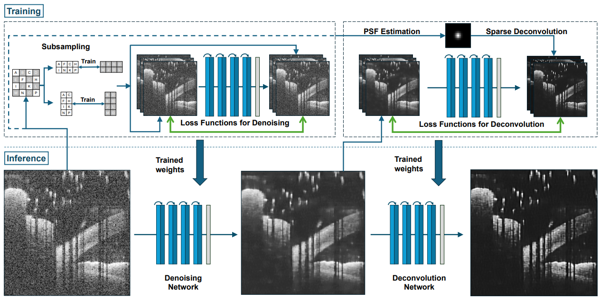
Schematic of the real-time OCT deconvolution framework, encompassing both training and inference phases. The training phase involves collecting a diverse dataset and applying a subsampling strategy to generate denoised images, which are then utilized to train a denoising network as detailed in this paper. Subsequently, the sparse deconvolution is applied to the denoised images to create a set of enhanced images for supervision. The trained network is subsequently deployed in the inference phase for direct enhancement of OCT scans, facilitating real-time visualization
First author Weiyi Zhang, a member of the research team, stated: "Our self-supervised framework integrates three core modules—denoising preprocessing, blind PSF estimation, and sparse deconvolution—to achieve real-time deconvolution relying solely on raw OCT images. Experimental validation demonstrates that this method improves axial and lateral resolution by approximately 40% compared to raw images, while effectively suppressing noise sidelobes generated by traditional Lucy-Richardson algorithms. For in vivo imaging scenarios such as ocular and vascular tissues, the method significantly enhances structural signal-to-noise ratio and the resolvability of tissue details. The lightweight network, containing only 69,000 parameters, processes kilopixel B-scan images in milliseconds on standard GPUs, meeting real-time requirements for clinical and industrial applications. The method also maintains stable performance in cross-dataset tests spanning different wavelengths (840/1060/1310 nm) and imaging modes (free-space/endoscopic), providing a registration-free solution for real-time OCT image enhancement."
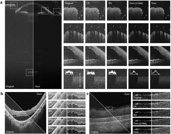
Enhanced OCT images of ocular samples using different deconvolution methods. (a) Comparison of human eye images, with insets showing detailed regions. (b) and (c) Posterior human eye and rabbit retina images before and after enhancement with our proposed method, highlighting improved clarity and resolution. LR, Lucy–Richardson deconvolution; SD, sparse deconvolution.
III. Future Perspectives
The research team plans to conduct in-depth investigations into the limitations of the current framework, with a focus on optimizing PSF modeling and dynamic estimation methods under non-ideal imaging conditions, and exploring physics-informed deformable convolutional network architectures to enhance generalization capabilities for complex samples. At the application level, the method will be extended to functional OCT imaging (e.g., hemodynamic analysis and polarization-sensitive OCT) to establish multimodal joint deconvolution models that preserve optical phase information. Concurrently, the team will develop a cross-device, cross-band standardized validation framework, design adaptive network compression algorithms to meet real-time processing demands in embedded OCT systems, and advance the translational application of this technology in clinical scenarios such as ophthalmic diagnostics and cardiovascular interventions through industry-academia-research collaborations.