


Methods for the photographic recording of ultrahigh-speed events are essential tools across many scientific disciplines. In contrast to traditional stroboscopic or pump-probe methods that require the repeatability of phenomena under study, "single shot" imaging techniques enable the study of non-repeatable ultrahigh-speed events. As a core approach to single-shot imaging, mapping photography/microscopy extends the frame rates of conventional image sensors through mechanisms of time-to-space optical mapping that assigns independent temporal frames to separate spatial positions that are recorded by one or more two-dimensional (2D) sensors. Among available techniques, optical diffraction has recently been implemented by our group as a nanosecond time gate for mapping photography based on the intrinsic dynamics of digital micromirror device (DMD) inter-pattern transition. This technique, termed diffraction-gated real-time ultrahigh-speed mapping (DRUM) photography, offers advantages in light throughput, cost efficiency, and passive operation, but has so far only been applied to intensity-based contrast imaging using temporal information distributed along one spatial axis in the sensor plane.
Building on the same system architecture as DRUM photography, we now present diffraction-gated real-time ultrahigh-speed mapping schlieren (DRUMS) photography for single-shot phase-gradient-contrast-based imaging at million-frame-per-second (Mfps) speeds. DRUMS photography presents two key innovations. First, by taking advantage of the DMD's role as a controllable spatial filter located at the optical Fourier plane of input scenes (Fig. 1a), DRUMS photography enables sensitivity to schlieren phenomena via the "knife-edge" filtering of spatial frequencies that are blocked by the selective isolation of DMD micromirrors during inter-pattern transition (Fig. 1b). Second, by incorporating additional temporal information distributed in off-axis diffraction orders, DRUMS photography significantly advances the core capabilities of DRUM photography, delivering a roughly two-fold increase to both frame rate and sequence depth (Fig. 1c).
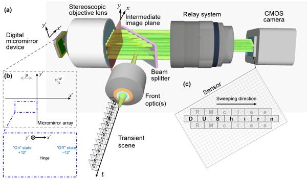
Fig. 1 Schematic of DRUMS photography. (a) Schematic of the system. (b) Schematic of the DMD micromirror array, regarded as the combination of two rectangular grids of pitch highlighted by the yellow and gray colors. The inset illustrates the flipping action of the DMD micromirrors during pattern transition, with the hinge axis oriented parallel to the -axis. (c) Illustrative example of how off-axis diffraction orders expand the temporal information recovered by the sensor.
The DRUMS photography technique was effectively utilized to investigate the interaction between femtosecond laser pulses and liquid water. As illustrated in Fig. 2(a), a femtosecond laser produced a single pump pulse (wavelength 1035 nm, pulse duration 350 fs, pulse energy 7 µJ), which was focused into distilled water contained in a glass cuvette via a lens with a focal length of 15 mm. The focused beam induced ionization of the distilled water, resulting in the formation of a plasma channel at the focal point. Figure 2(b) presents five representative frame images capturing the refractive index perturbations caused by laser-induced breakdown. Furthermore, Fig. 2(c) illustrates the temporal evolution of the plasma channel length. According to the data, the channel length was measured to be 434 µm at 0.23 µs and subsequently decreased to 70 µm at 1.23 µs.
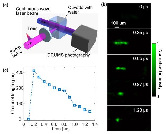
Fig. 2 DRUMS photography of laser-induced breakdown in distilled water. (a) Schematic of the experimental setup. (b) Selected DRUMS photography frames showing the evolution of the laser induced plasma channel in distilled water using a 7 µJ pump pulse. (c) Time history of the channel length.
DRUMS photography, a single-shot 2D ultrahigh-speed schlieren imaging technique, achieves a frame rate of 9.8 Mfps with a sequence depth of 13 frames. This technique has been utilized to investigate laser-liquid interactions and holds promise for studying the impact of tissue heterogeneity on laser ablation in minimally invasive surgeries. The work, titled "Ultrahigh-speed schlieren photography via diffraction-gated real-time mapping" and published in Advanced Imaging (2025, Vol. 2, Issue 1), demonstrates substantial advancements in ultra-high-speed schlieren imaging methodologies.