


 View fulltext
Advanced Photonics
Co-Editors-in-Chief
Xiao-Cong (Larry) Yuan, Anatoly Zayats
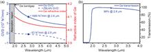
Zhipeng Qin, Guoqiang Xie, Hongan Gu, Ting Hai, Peng Yuan, Jingui Ma, and Liejia Qian
The mode-locked fluoride fiber laser (MLFFL) is an exciting platform for directly generating ultrashort pulses in the mid-infrared (mid-IR). However, owing to difficulty in managing the dispersion in fluoride fiber lasers, MLFFLs are restricted to the soliton regime, hindering pulse-energy scaling. We overcame the problem of dispersion management by utilizing the huge normal dispersion generated near the absorption edge of an infrared-bandgap semiconductor and promoted MLFFL from soliton to breathing-pulse mode-locking. In the breathing-pulse regime, the accumulated nonlinear phase shift can be significantly reduced in the cavity, and the pulse-energy-limitation effect is mitigated. The breathing-pulse MLFFL directly produced a pulse energy of 9.3 nJ and pulse duration of 215 fs, with a record peak power of 43.3 kW at 2.8 μm. Our work paves the way for the pulse-energy and peak-power scaling of mid-IR fluoride fiber lasers, enabling a wide range of applications.
Dec. 27, 2019Vol. 1 Issue 6 065001 (2019)
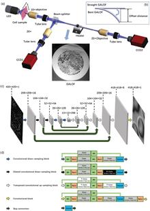
Jian Zhao, Yangyang Sun, Hongbo Zhu, Zheyuan Zhu, Jose E. Antonio-Lopez, Rodrigo Amezcua Correa, Shuo Pang, and Axel Schulzgen
We demonstrate a deep-learning-based fiber imaging system that can transfer real-time artifact-free cell images through a meter-long Anderson localizing optical fiber. The cell samples are illuminated by an incoherent LED light source. A deep convolutional neural network is applied to the image reconstruction process. The network training uses data generated by a setup with straight fiber at room temperature (~20 ° C) but can be utilized directly for high-fidelity reconstruction of cell images that are transported through fiber with a few degrees bend or fiber with segments heated up to 50°C. In addition, cell images located several millimeters away from the bare fiber end can be transported and recovered successfully without the assistance of distal optics. We provide evidence that the trained neural network is able to transfer its learning to recover images of cells featuring very different morphologies and classes that are never “seen” during the training process.
Nov. 11, 2019Vol. 1 Issue 6 066001 (2019)
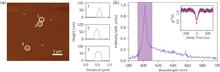
Minh Nguyen, Niko Nikolay, Carlo Bradac, Mehran Kianinia, Evgeny A. Ekimov, Noah Mendelson, Oliver Benson, and Igor Aharonovich
Color centers in diamond—especially group IV defects—have been advanced as a viable solid-state platform for quantum photonics and information technologies. We investigate the photodynamics and characteristics of germanium-vacancy (GeV) centers hosted in high-pressure high-temperature diamond nanocrystals. Through back-focal plane imaging, we analyze the far-field radiation pattern of the investigated emitters and derive a crossed-dipole emission, which is strongly aligned along one axis. We use this information in combination with lifetime measurements to extract the decay rate statistics of the GeV emitters and determine their quantum efficiency, which we estimated to be ~ ( 22 ± 2 ) % . Our results offer further insight into the photodynamic properties of the GeV center in nanodiamonds and confirm its suitability as a desirable system for quantum technologies.
Dec. 26, 2019Vol. 1 Issue 6 066002 (2019)
Shimon Rubin, and Yeshaiahu Fainman
Optical metamaterials and metasurfaces, which emerged in the course of the last few decades, have revolutionized our understanding of light and light–matter interaction. While solid materials are naturally employed as key building elements for construction of optical metamaterials mainly due to their structural stability, practically no attention was given to study of liquid-made optical two-dimensional (2-D) metasurfaces and the underlying interaction regimes between surface optical modes and liquids. We theoretically demonstrate that surface plasmon polaritons and slab waveguide modes that propagate within a thin liquid dielectric film trigger optical self-induced interaction facilitated by surface tension effects, which leads to the formation of 2-D optical liquid-made lattices/metasurfaces with tunable symmetry and can be leveraged for tuning of lasing modes. Furthermore, we show that the symmetry breaking of the 2-D optical liquid lattice leads to phase transition and tuning of its topological properties, which allows the formation, destruction, and movement of Dirac-points in the k-space. Our results indicate that optical liquid lattices support extremely low lasing threshold relative to solid dielectric films and have the potential to serve as configurable analogous computation platform.
Dec. 27, 2019Vol. 1 Issue 6 066003 (2019)
Jiaji Li, Alex Matlock, Yunzhe Li, Qian Chen, Chao Zuo, and Lei Tian
We demonstrate a label-free, scan-free intensity diffraction tomography technique utilizing annular illumination (aIDT) to rapidly characterize large-volume three-dimensional (3-D) refractive index distributions in vitro. By optimally matching the illumination geometry to the microscope pupil, our technique reduces the data requirement by 60 times to achieve high-speed 10-Hz volume rates. Using eight intensity images, we recover volumes of ~350 μm × 100 μm × 20 μm, with near diffraction-limited lateral resolution of ~ 487 nm and axial resolution of ~ 3.4 μm. The attained large volume rate and high-resolution enable 3-D quantitative phase imaging of complex living biological samples across multiple length scales. We demonstrate aIDT’s capabilities on unicellular diatom microalgae, epithelial buccal cell clusters with native bacteria, and live Caenorhabditis elegans specimens. Within these samples, we recover macroscale cellular structures, subcellular organelles, and dynamic micro-organism tissues with minimal motion artifacts. Quantifying such features has significant utility in oncology, immunology, and cellular pathophysiology, where these morphological features are evaluated for changes in the presence of disease, parasites, and new drug treatments. Finally, we simulate the aIDT system to highlight the accuracy and sensitivity of the proposed technique. aIDT shows promise as a powerful high-speed, label-free computational microscopy approach for applications where natural imaging is required to evaluate environmental effects on a sample in real time.
Dec. 28, 2019Vol. 1 Issue 6 066004 (2019)
© Copyright 2018-2021 | Chinese Laser Press.
All Rights Reserved 沪ICP备15018463号-20