


Please enter the answer below before you can view the full text.

Jiaqing Tao, Zexi Zheng, Huazhong Xiang, and Xianyang Tian
ObjectiveImage-based non-contact measurements for pulse wave remote acquisition and monitoring have an important practical value in clinical use. Accurate pulse waves are a major prerequisite for measuring parameters of human physiology such as the heart rate, heart rate variability, blood oxygen concentration, and blood pressure. Based on the fact that the carotid artery is the closest observable artery to the human heart and contains a wealth of physiological information, vibrations of the epidermis caused by blood flow can be observed on the surface of the human body. In addition, the amplitude of random motion on the neck is much smaller than that on the human face. Accordingly, the signal source is set on the neck for better observations, less disturbance, and more up-to-date results. Under normal circumstances, the pulsation of the human carotid artery causes a small vibration that is visible to the naked eye and can be obtained by analyzing the vibration using conventional image and signal processing methods. However, in clinical practice, some patients have a relatively weak carotid pulse, and the existing statistical signal processing and time-frequency domain signal processing methods are inadequate for obtaining the desired signal. Thus, a new signal processing method is required for these types of situations.MethodUnder the illumination of an 850 nm near-infrared light source, a near-infrared camera was used to continuously shoot the image sequence of the vibration of the neck skin. The final signal was obtained through a series of images and signal processing. The specific process is described as follows. First, the region of interest (ROI) was obtained using the inter-frame difference method. The original gray signal was then obtained by calculating the mean value of the ROI. Next, the original signal was normalized from the gray signal at an interval of 0 to 1. Finally, the desired pulse wave signal was acquired using bandpass filtering and the proposed multi-region dominant frequency enhancement (MRDFE) method. The MRDFE method is a joint algorithm that combines frequency domain processing and principal component analysis in two steps. In the first step, the signal obtained in each ROI was assigned the weight of the dominant frequency signal-to-noise ratio. In the second step, the signals in these ROI channels were evaluated by principal component analysis, and the feature vector corresponding to the first eigenvalue obtained was the final output signal. To further demonstrate the robustness of the algorithm, we established our own database, which contained 24 sets of weak pulse vibration image sequences. In dealing with these data, we compared our method with other existing algorithms based on four indicators: periodic integrity, periodic variation, tidal wave integrity, and repulse wave integrity.Results and DiscussionsThe proposed MRDFE method can be used to obtain pulse waves with preserved feature points in a weak pulse situation (Fig. 4). To compare the MRDFE method with other conventional methods, a feature point recognition algorithm called the stepwise threshold descent method was used to detect feature points from the final signal obtained by each method. Our experimental results show that the proposed method performs much better than the other three conventional algorithms. Our method exhibits a more stable periodic state and retains approximately 70% of the tidal wave characteristics and more than 50% of the repulse wave characteristics (Table 1). Based on observations of the signals derived from the different methods (Fig. 7), the periodicity of the pulse wave obtained by our method is more obvious, and more feature points are preserved. The MRDFE method enhances the signal with a high signal-to-noise ratio and weakens the signal with a low signal-to-noise ratio through weight assignment, yielding satisfactory results.ConclusionsThis study presents a method for obtaining pulse wave signals under the condition of weak pulse vibrations of the carotid artery. With a near-infrared light source used for illumination, the image sequence of neck skin vibration was captured by a camera. Several ROIs were selected from the image sequence, and the initial signal was acquired using outlier processing and bandpass filtering. The pulse wave signal of the weak pulse vibration was then processed successfully using the MRDFE method. Compared with other signal processing methods, the analytical results show that the signal obtained by the MRDFE method is of higher quality, preserves a greater number of feature points, and provides better cycle integrity. Our analysis and experimental results show that the proposed method is superior in performance to the existing signal processing methods. Robust and reliable pulse wave signals can be obtained using this method and applied in further measurements of the heart rate, heart rate variability, blood oxygen, and even blood pressure. The MRDFE method adds considerable value to new signal processing for image-based non-contact pulse wave extraction.
May. 10, 2023Vol. 50 Issue 9 0907101 (2023)
Li Zhao, Ri Zhou, Guannan Liu, Guishan Peng, Chenguang Wang, Xiaoteng Jia, and Geyu Lu
ObjectiveLipid droplets are important organelles closely associated with various cellular physiological activities. Confocal fluorescence imaging is a powerful tool for observing lipid droplets and studying their diverse functions. However, lipid droplet fluorescent probes with the high fluorescence intensity and labeling selectivity required for cellular lipid droplet fluorescence imaging are limited, severely limiting the in-depth study of lipid droplets. In this study, we develop Lipi-QL, a quinoline-derivative lipid droplet fluorescent probe with fluorescence-switching properties.MethodsThe probe exhibits high selectivity for lipid droplet labeling owing to its sensitive polar quenching fluorescence properties. The donor-type molecular structure also confers high fluorescence intensity and large Stokes shifts on the probe. When using this probe for confocal fluorescence imaging of cellular lipid droplets, significantly better labeling selectivity is achieved at varying concentrations than when using the commercial BODIPY 493/503 lipid droplet probe. Additionally, three-dimensional confocal imaging of fixed cells and four-color confocal imaging of live cells are performed using this fluorescent probe. The development of this probe provides a powerful tool for studying the physiological functions of lipid droplets and provides a new idea for the design of new highly labeled selective fluorescent probes.Results and DiscussionsAs shown in Fig.1(c), the probe exhibits highly efficient fluorescence emission when the water volume fraction is 0, indicating that it can exhibit high fluorescence intensity within lipid droplets. When the water volume fraction gradually increases, the probe exhibits extremely sensitive fluorescence quenching properties: quenching most of the fluorescence emission when the water volume fraction is only 1%. When the water volume fraction increases to 20%, the probe's emission is almost completely quenched, and the fluorescence signal disappears. This indicates that even if a small portion of the probe enters the cell and stains organelles other than lipid droplets, the fluorescence emission is quenched by the polar environment in which it is placed, thus showing a high selectivity for lipid droplet staining. We also test the fluorescence switching characteristics of the commercial lipid droplet dye, BODIPY 493/503. As shown in Fig.1(d), the fluorescence quenching of BODIPY 493/503 in the dioxane solution with 40% water volume fraction is not apparent, which may be the main reason for its poor lipid droplet staining selectivity. Figure 3 shows that the Lipi-QL fluorescent probe efficiently stains cellular lipid droplets at different concentrations. In contrast, BODIPY 493/503 stains lipid droplets much less selectively, staining other membrane-like cellular structures in addition to cellular lipid droplets with a lower imaging signal-to-noise ratio. This staining selectivity comparison highlights the significant advantage of the polar quenching luminescence property of the Lipi-QL fluorescent probe for the efficient and selective labeling of cellular lipid droplets. After washing the free probe with phosphate buffered saline (PBS), three-dimensional confocal imaging is performed. The experiment is performed at a high xy-plane point resolution with a small z-sweep step (200 nm) to obtain high-quality 3D confocal photographs (Fig.4). The spatial distribution of intracellular lipid droplets can be seen clearly in this photograph, demonstrating the usefulness of the probe for 3D confocal imaging. The Lipi-QL fluorescent probe is also used for multicolor confocal imaging because of its excellent performance. The nuclei, lipid droplets, lysosomes, and mitochondria of live HeLa cells are stained with the Hoechst 33342 commercial dye for nuclei, Lipi-QL commercial dye for lipid droplets, LysoTracker Deep Red commercial dye for lysosomes, and MitoTracker Deep Red commercial dye for mitochondria, respectively. High-quality four-color confocal images of living cells are successfully obtained by performing confocal fluorescence. Based on the different absorption and emission spectra of these four fluorescent probes, imaging is performed through line-by-line scanning, effectively avoiding the occurrence of crosstalk between individual fluorescent channels.ConclusionsIn conclusion, an advanced lipid droplet fluorescent probe with fluorescence switching properties, Lipi-QL, is developed in this study, which allows for the efficient and selective labeling of cellular lipid droplets. The probe also has high fluorescence brightness, a large Stokes shift, and good biocompatibility. Based on these excellent properties, high-quality three-dimensional confocal imaging of fixed cells and four-color confocal imaging of live cells are successfully achieved using this probe, highlighting its utility in lipid droplet fluorescence imaging. The development of this probe provides an effective tool for cell biology studies of lipid droplets and a new approach for the design and synthesis of highly labeled selective fluorescent probes.
May. 10, 2023Vol. 50 Issue 9 0907102 (2023)
Xiaolong Chen, Yizhi Liang, Xiaoxuan Zhong, Xue Bai, Long Jin, Wei Huang, Cheng Huang, Xiaobing Niu, Shanshan Guo, and Baiou Guan
ObjectiveMicrocirculatory dysfunction may cause circulatory failure, insufficient oxygen delivery, and fatal risks. Microscopes are used to observe microcirculation, but they can only image superficial tissues. In addition, they can hardly provide functional information. In this study, we report a photoacoustic endoscope for in vivo imaging of the gastrointestinal microcirculation. The imaging probe is inserted into the rectum of a small animal for rotational-scanning endoscopic imaging. The vascular structures in the gastrointestinal wall can be visualized by detecting the ultrasound excited by the pulsed laser. Moreover, the blood oxygen saturation can be measured and imaged with a dual-wavelength excitation based on the difference in the optical absorption spectrum between oxy- and deoxygenated hemoglobin. We believe that this technology is capable of detecting the functional changes associated with microcirculation diseases with minimal invasion.MethodsThe imaging system consists of an endoscopic imaging probe, dual-wavelength pulsed laser source, rotary scanning device, and data acquisition and control module. First, we design an all-fiber endoscope probe containing two functional optical fibers as follows: one responsible for guiding and focusing the pulsed light and the other equipped with a laser ultrasonic sensor to detect the photoacoustic signal. Second, we design a rotational scanning device that rotates synchronously with the probe to achieve fast and unidirectional rotary scanning. This is achieved by miniaturizing the 980-nm pump laser, the optical amplifier, and the photodetector. Finally, we perform high-resolution in vivo endoscopic imaging of the rat rectum.Results and DiscussionsThe endoscope probe has a diameter of 2.75 mm, a resolution of 12.5 μm, signal jitter root mean square of 2.5%, and a B-scan frequency of 1 Hz. The instrument is stable and provides spatial resolution in high-speed scanning and is suitable for small animal digestive tract endoscopic imaging. The functional imaging results of the rectum of healthy rats show that we achieve 360°scanning, obtain the three-dimensional imaging results of hemoglobin concentration distribution, and show the vascular structure of the inner wall of the rat rectum. Along with the spatial distribution of blood oxygen saturation, the images show the distributions of the artery and vein in the inner wall (Fig. 3). The imaging results of septic rats show the changes in microcirculation. According to the imaging result, the number of blood vessels in the intestine of rats gradually decreases, and the blood oxygen saturation also declines in 5 h (Fig. 4). The above results reflect the phenomenon of insufficient tissue perfusion caused by sepsis.ConclusionsIn summary, we develop a photoacoustic endoscope for in vivo rectal imaging. By using fiber optic ultrasound sensors, the endoscope can image the vascular structure and visualize the changes in oxygen saturation. By using this endoscope, we can visualize the gastrointestinal microcirculatory disorder caused by lesions. The structure, number, and blood oxygen saturation level of blood vessels are significantly changed. The experimental results show that this technology can provide functional imaging results with high spatial resolution and high contrast in the endoscopic imaging of narrow cavity structures, thus providing a feasible imaging method for the characterization of microcirculation status and the diagnosis and treatment of acute and severe diseases.
May. 10, 2023Vol. 50 Issue 9 0907103 (2023)
Zhiguo Shi, Yang Zhang, Quanfu Wang, Zhongsheng Li, Xia Wang, Meili Dong, Jingshu Ni, Yao Huang, Shengzhao Zhang, Yikun Wang, and Yuanzhi Zhang
ObjectiveThe Muller matrix, as a method for characterizing the polarization properties of samples, contains complete information about the polarization properties of samples, and it has become an important indicator for characterizing pathological tissues in basic and preclinical studies. However, in the traditional polarized light imaging method for measuring the Muller matrix, the scattering depth of polarized light in collagen tissue cannot be controlled. The obtained Muller matrix information is the average of unknown depths in collagen tissue, and it is impossible to accurately measure the Muller matrix information of the pathological tissue area. Polarized spatial frequency domain imaging (PSFDI), which combines spatial frequency domain imaging (SFDI) and polarized light imaging, is applied to measure the optical properties of biological tissues accurately.MethodsSFDI relates the spatial frequency of the projected stripe pattern to the penetration depth of the detected light, and the imaging depth can be controlled by controlling the spatial frequency of the projected light. We designed and validated a polarization SFDI system that uses the SFDI technique to control the imaging depth, projects the streak pattern onto the surface of the measured tissue, constructs a polarizer and detector to modulate the polarization state of the polarized light, and then acquires the image data using a CMOS camera and calculates the Mueller matrix information.Results and DiscussionsExperimental results showed that the grey-scale plate diffuse reflectance measured by the polarization SFDI system was linearly correlated with the standard value (R2=0.99988). The depolarization coefficient tends to be proportional to the fat emulsion volume fraction, the two-way attenuation coefficient increases with the increase of two-way attenuation owing to the two-way attenuator, and the accurate measurement of the phase delay of the quarter-wave and full-wave plates indicates that the system can accurately measure the sample polarization parameters. A comparison of uniform light field illumination and polarization-sensitive SFDI shows that the latter effectively controls the depth and accurately measures the shallow Mueller matrix of the sample. The results of this study are expected to effectively improve the accuracy of the detection of polarization characteristics of superficial tissues and promote early tumor detection.ConclusionsIn this study, a PSFDI system is developed based on polarized light imaging and SFDI, and the device structure, measurement method, and data processing method are introduced. By performing the error calibration of the PSFDI device, the measurement error of the device can be less than 2%. By performing Mueller matrix imaging on tissues, we verified the reliability of the device to measure tissue polarization Mueller and the accuracy of the Mueller matrix decomposition; hence, PSFDI can be used to obtain the optical properties of various samples. The PSFDI can accurately image pathological regions, providing accurate physiological parameters for pathological analysis, and it has a wide range of biomedical applications.
May. 10, 2023Vol. 50 Issue 9 0907104 (2023)
Yanhong Ma, and Pengfei Zhang
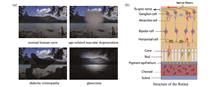
SignificanceRetinal opto-physiology is the physiological response of the retina to a visible light stimulus, reflecting the function of the retina to a certain extent. Optoretinography, also termed optoretinogram (ORG), is a newly developed functional imaging technique for precisely quantifying the opto-physiological response of the retina. It uses optical coherence tomography combined with controllable light stimulus, to accurately measure retinal morphology and optical property changes in response to a light stimulus by detecting the peak position and scattering intensity alternations in optical coherence tomography (OCT) images. Moreover, ORG can achieve microscale lateral resolution, nanoscale axial resolution, and millisecond temporal resolution by combining adaptive optics and phase analysis techniques, and be used to measure opto-physiological functions of the retina at the cellular level. To perform ORG, only an optical stimulus unit is required to be added to the existing OCT system. Currently, it is still in the stage of technology development and mechanism exploration. Once standards are established, ORG may be used in broad ophthalmic research and clinical practice. In this study, the history of OCT functional imaging development for probing the retinal opto-physiological signal is systematically reviewed, the latest progress in ORG technology is summarized, and several future directions of ORG technology are discussed.ProgressBecause of the excellent spatial resolution of OCT and its extensive use in basic research, clinical diagnosis, and treatment, researchers have been committed to capturing the functional response of nerve cells to a visible light stimulus using OCT. Early studies of OCT photo-physiological functional imaging focused on finding the changes in retinal scattering signals after light stimulation. With the gradual improvement of OCT performance (such as resolution and imaging speed) and the wide application of in vivo imaging technology, a series of breakthroughs have been made in the field of OCT retinal functional imaging in recent years. The optical path difference changes of cone outer segment in living human retina after a light stimulus [Fig. 2(a)-(c)] were successfully measured using high-speed full-field OCT combined with phase analysis technology. Subsequently, Zhang et al. used OCT to observe the function signals of mouse retina in response to the light stimulation with different intensities. They reported the changes in the thickness of the rod outer segment at a micron level and changes in scattering signal intensities of several retinal layers [Fig. 2(d)-(h)]. Moreover, they confirmed that the response signals came from the visual photo-transduction process using gene knockout mice. Furthermore, due to the advantages of adaptive optics enhanced OCT (AO-OCT) in imaging resolution, researchers have successfully measured the photo-physiological signal at the level of a single cone cell in the human eye. For example, Zhang et al. measured the functional response of human cone cells after light stimulation using AO-OCT in 2019 and successfully distinguished three types of cone cells in the human eyes via different cone cell responses to different color stimuli (Fig. 3). In the recent studies, several experimental groups have conducted the diseases or mechanism research on the retina from a physiological perspective. Qian et al. changed the mouse retina through transgenic or drug methods to carry out controlled experiments with conventional wild mice. They found that changes in the thickness of the mouse’s external retina after light stimulation were affected by the base level of mitochondrial respiration and oxidative stress reaction. This suggests the favorable conditions for the clinical application of OCT-based photo-physiological functional imaging.Conclusions and ProspectsThe method based on OCT retinal opto-physiological functional imaging and visible light stimulation is collectively called optoretinogram. ORG is a new technology in which the obtained opto-physiological signals can reflect the function of retinal tissue. This addresses the limitation of conventional OCT imaging technology which can only provide details of the retinal structure. Moreover, studies of human and animal retina indicate that retinal responses to light stimulation involve subtle changes in several structures including the rod, cone, retinal pigment epithelium, Bruch's membrane, and choroid. All are closely related to the structures affected by various retinal diseases. Thus, to some extent, early retinopathy may affect the intensity and change rate of retinal opto-physiological function signal. Therefore, this can provide a new diagnostic basis for detection of early disease by measuring the abnormal changes in the opto-physiological function signal.Future developments of ORG may include the following four aspects: 1) realizing the local analytical capability of ORG while maintaining wide-field macroscopic imaging; 2) further enhancement of the sensitivity of OCT to enable detection of weaker functional signals; 3) extracting the functional signal quickly and automatically and exploring the characteristic functional signal of early retinopathy; and 4) optimization and standardization of experimental methods.
May. 10, 2023Vol. 50 Issue 9 0907105 (2023)
Linjun Zhai, Yuqing Fu, and Yongzhao Du
SignificanceBlood flow is an important parameter for measuring vital signs, and hemodynamic parameters are functional indicators of the microcirculatory system of the skin, brain, heart, liver, kidneys, and other organs. Therefore, dynamic blood flow monitoring has important application value and significance in clinical and basic life science fields, such as clinical diagnosis, intraoperative guidance, drug research, disease mechanism research, and neuroscience. Laser speckle contrast imaging (LSCI) is a full-field optical imaging technique that uses the spatial and temporal statistical properties of laser scattering intensity to monitor the blood flow of tissues in vivo. It uses simple equipment, is non-invasive, and has a fast imaging speed and high spatial resolution. Additionally, it does not require the injection of a contrast agent and can perform continuous measures for a long time. Consequently, it is widely used to measure microcirculatory blood flow parameters such as the vessel diameter, blood flow velocity, blood perfusion, and blood density in tissues and organs. It also can help doctors locate the lesion precisely with clear and accurate blood flow data,and then analyze the corresponding functional response and pathological mechanisms, which has become one of the most important tools for the clinical diagnosis of fundus diseases, skin diseases, brain diseases, and so on. In addition, it is also an important tool for basic life science research in drugs, cardiovascular and cerebrovascular diseases, and brain cognitive and behavioral sciences. Consequently, in-depth research on novel LSCI techniques with high imaging quality is valuable and significant for improving the quality of medical care and promoting the development of basic research in life science.ProgressIn the past decades, many researchers have conducted extensive researches on how to improve the quality of LSCI and expand the scope of LSCI applications, and they have had positive progress. For example, a few research groups like Luo Qingming and Li Pengcheng at Huazhong University of Science and Technology, and Tong Shanbao at Shanghai Jiao Tong University have worked on portable LSCI systems, high signal-to-noise ratio LSCI, and high resolution LSCI, which have promoted the development of LSCI in China. Researchers abroad like Boas at Boston University, Zakharov at the University of Fribourg, and Dunn at the University of Texas at Austin have worked on high-precision imaging using LSCI techniques, such as static scattered light correction and quantitative analysis of LSCI, which has also greatly promoted the development of key techniques and novel LSCI applications.In this paper, we presented a systematic, comprehensive and integrated analysis, review and summary of the current researches about key techniques and applications of novel LSCI at home and abroad emphatically from the aspect of high signal-to-noise ratio LSCI, high-resolution LSCI, high-precision LSCI, large imaging depth LSCI, and novel LSCI systems based on the investigations of current literature. In this way, we can help researchers learn more about the frontier technologies of LSCI and understand the technical challenges we faced, and we can provide ideas with a reference value to promote the development of high-quality, highly practical, and innovative LSCI systems to meet the needs of clinical diagnosis and basic biomedical research. The review consisted of the following contents: First, the technical problems of measuring deep blood flow and achieving high resolution, high signal-to-noise ratio, and high precision have been systematically summarized, and the corresponding solutions are indicated. Subsequently, we review high signal-to-noise ratio LSCI techniques based on anisotropic filtering, eigenvalue-decomposition, and transformation domain collaborative filtering methods. Meanwhile, high-resolution LSCI for motion artifact, out-of-focus blur, and non-uniform light intensity correction are also summarized. Third, we elaborate the high-precision LSCI from the perspective of static scattered light correction, quantitative analysis, and novel LSCI algorithms. After summarizing the LSCI with a large imaging depth, we introduce the latest research on the novel LSCI system and its applications in the fields of cortical blood flow imaging, surgical and therapeutic procedures, and brain and cognitive-behavioral sciences. Finally, we discuss and look forward to the development of LSCI in the future.Conclusions and ProspectsIn conclusion, LSCI has made qualitative leaps and developments in theory, imaging systems, computational methods, and clinical applications. The imaging quality of LSCI has been developed to have a high signal-to-noise ratio, high resolution, high accuracy, and large imaging depth. However, as the application scenarios of LSCI become more and more complex, which introduces greater challenges to the development of key techniques and application of LSCI. In the future, LSCI will be deeply integrated with emerging interdisciplinary fields such as biomedicine, optoelectronic information, artificial intelligence, and big data. In addition, new breakthroughs are expected in the following respects. (1) Quantitative analysis capacity. The capacity is still an important fundamental issue for LSCI in functional applications. (2) Combination of LSCI with new endoscopic technology (this will enable the noninvasive measurement of blood flow). (3) Miniaturization and integration. The development of new materials and electronic devices will certainly promote the miniaturization and integration of new LSCI systems. (4) Combination of LSCI with artificial intelligence. Artificial intelligence will further promote the development of LSCI technologies and their applications. (5) Combination with other imaging modalities (this will build a new model for LSCI-based multimodal clinical diagnostic applications). It is believed that LSCI will show a synergistic development trend in the future. We look forward to seeing the development of technologies and applications of LSCI.
May. 10, 2023Vol. 50 Issue 9 0907106 (2023)
Chijian Li, Jing Yao, Yufeng Gao, Puxiang Lai, Yuezhi He, Sumin Qi, and Wei Zheng
ObjectiveTwo-photon microscopy (TPM) imaging has been widely used in many fields, such as in vivo tumor imaging, neuroimaging, and brain disease research. However, the small field-of-view (FOV) in two-photon imaging (typically within diameter of 1 mm) limits its further application. Although the FOV can be extended through adaptive optics technology, the complex optical paths, additional device costs, and cumbersome operating procedures limit its promotion. In this study, we propose the use of deep learning technology instead of adaptive optics technology to expand the FOV of two-photon imaging. The large FOV of TPM can be realized without additional hardware (such as a special objective lens or phase compensation device). In addition, a BN-free attention activation residual U-Net (nBRAnet) network framework is designed for this imaging method, which can efficiently correct aberrations without requiring wavefront detection.MethodsCommercially available objectives have a nominal imaging FOV that has been calibrated by the manufacturer. Within the nominal FOV, the objective lens exhibits negligible aberrations. However, the aberrations increase dramatically beyond the nominal FOV. Therefore, the imaging FOV of the objective lens is limited to its nominal FOV. In this study, we improved the imaging quality of the FOV outside the nominal region by combining adaptive optics (AO) and deep learning. Aberrant and AO-corrected images were collected outside the nominal FOV. Thus, we obtained a paired dataset consisting of AO-corrected and uncorrected images. A supervised neural network was trained using the aberrated images as the input and the AO-corrected images as the output. After training, the images collected from regions outside the nominal FOV could be fed directly to the network. Aberration-corrected images were produced, and the imaging system could be used without AO hardware.Results and DiscussionsThe experimental test results include the imaging results of samples such as fluorescent beads with diameter of 1 μm and Thy1-GFP and CX3CR1-GFP mouse brain slices, and the results of the corresponding network output. The high peak signal-to-noise ratio (PSNR) values of the test output and ground truth demonstrate the feasibility of extending the FOV to TPM imaging using deep learning. At the same time, the intensity contrast between the nBRAnet network output image and the ground truth on the horizontal line is compared in detail (Figs. 3, 4, and 5). The extended FOV of different samples is randomly selected for analysis, and a high degree of coincidence is observed in the intensity comparison. The experimental results show that after using the network, both the resolution and fluorescence intensity can be restored to a level where there is almost no aberration, which is close to the result after correcting using AO hardware. To demonstrate the advantages of the network framework designed in this study, the traditional U-Net structure and the very deep super-resolution (VDSR) model are used to compare with ours. When using the same training dataset to train different models, we find that the experimental results of the VDSR model contain a considerable amount of noise, whereas the experimental results of the U-Net network lose some details (Fig. 6). The high PSNR values clearly demonstrate the strength of our nBRAnet network framework (Table 3).ConclusionsThis study provides a novel method to effectively extend the FOV of TPM imaging by designing an nBRAnet network framework. In other words, deep learning was used to enhance the acquired image and expand the nominal FOV for commercial objectives. The experimental results show that images from extended FOVs can be restored to their AO-corrected versions using the trained network. That is, deep learning technology could be used instead of AO hardware technology to expand the FOV of commercially available objectives. This simplifies the operation and reduces the system cost. An extended FOV obtained using deep learning can be employed for cross-regional or whole-brain imaging.
May. 10, 2023Vol. 50 Issue 9 0907107 (2023)
Lijing Zhang, Binbin Wang, Wei Wang, Bo Wu, and Nan Zhang
ObjectiveIn surgical navigation system-assisted pedicle screw fixation, preoperative and intraoperative point clouds registration accuracy is crucial for positioning and navigation. When the patient's preoperative space is accurately registered to the actual surgical space, the surgical instrument can be guided to the patient's surgical site, and the planned surgical path can be accurately implemented during the operation. The preoperative point cloud is obtained by reconstructing the patients' preoperative CT, while a structural light scanner obtains the intraoperative point cloud during the operation. The acquisition methods of two-point clouds differ; hence, their densities and initial poses are quite different. Therefore, they are cross-source point clouds. Moreover, the scanned intraoperative point cloud is a small part of an entire spine since the exposed spine is very limited during an operation. Hence, the overlapping ratio of the preoperative and intraoperative point clouds is low. Existing registration algorithms are prone to fail or derive a low accuracy in preoperative and intraoperative point cloud registration. To solve these problems, this study proposes a preoperative and intraoperative point clouds registration algorithm with cross-source and a small overlapping ratio for pedicle screw fixation.MethodsThis study proposes a preoperative and intraoperative point clouds registration algorithm with cross-source and low overlapping ratio based on Farthest Point Sampling (FPS). The proposed algorithm includes coarse and fine registration. The coarse registration comprises three steps. Firstly, the voxel filter was used to down-sample the intraoperative point cloud to bring its density close to the preoperative point cloud. Secondly, the Fast Point Feature Histogram (FPFH) features of the intraoperative point cloud were extracted. The FPS was used to sample the preoperative point cloud, and then the preoperative point cloud was divided into several local regions by kd tree algorithm. These local regions formed the candidate set. Thirdly, the candidate set was traversed to calculate the FPFH features of each local region. The Sample Consensus Initial Alignment (SAC-IA) feature matching method was to realize feature matching and pose transformation estimation of intraoperative point cloud. The distance errors deduced by the SAC-IA method between the intraoperative point cloud and local point cloud were compared and the local region with the minimum distance error was selected as the optimal local region. The transformation of the optimal local region was the intraoperative and preoperative points clouds coarse registration's transformation. In fine registration, the Iterative Closest Point (ICP) algorithm was adopted to further align the intraoperative and the preoperative point cloud. It is performed based on the coarse registration result. The optimal local region is used as the target point cloud at this stage. Using the FPS and SAC-IC methods, an optimal local region sampled from the preoperative point cloud is obtained, enabling the point clouds can align under large original pose difference and low overlapping ratio conditions. In fine registration, the ICP algorithm was adopted to further align the intraoperative and the preoperative point clouds. The fine registration was performed based on the coarse registration results. The optimal local region was used as the target point cloud at this stage.Results and DiscussionsBased on the FPS method, the preoperative point cloud is divided into several local regions (Fig. 2). An optimal local neighborhood is derived to complete registration with the intraoperative point cloud. This study adopts nine pairs of preoperative and intraoperative point clouds for testing, with different overlapping ratios and initial poses (Table 1). The visualization results of the registration process using the proposed algorithm are shown in Fig. 3, including the initial pose of nine pairs of point clouds, the sampling results of FPS, and the coarse and fine registration results. To evaluate the performance of the proposed algorithm, two state-of-the-art registration algorithms, FPFH+ICP and SHOT+ICP, are adopted for comparison (Table 2). The proposed algorithm achieves the minimum coarse registration error, providing better alignment for ICP fine registration. A comparison of the final registration transformation matrix with the ground truth shows that the average rotation error is 0.406° and the translation error is 0.474 mm, which meets clinical requirements. Simultaneously, the registration time of the proposed algorithm is less than 2 min, which is adequate for an operation. In addition, the registration success rates of the three algorithms in the experiment are compared (Table 3). The successfully registered FPFH+ICP and SHOT+ICP algorithm samples are 6 out of 9. While for the proposed algorithm, it is 9 out of 9, demonstrating that the registration success rate increased from 66.67% to 100%.ConclusionsThis study proposes a point cloud registration algorithm with cross-source and a low overlapping ratio for pedicle screw fixation. Through the registration of preoperative and intraoperative point clouds of a lumbar vertebra with an overlapping ratio of less than 3%, the experimental results show that the proposed algorithm based on FPS can resolve the problems of density differences, large initial pose differences, and low overlapping ratios in the preoperative and intraoperative point clouds registration of pedicle screw fixation assisted by a surgical navigation system. High precision registration can be realized, improving the accuracy and safety of surgical navigation systems. The research only considers the rigid transformation of preoperative and intraoperative point clouds. Preoperative and intraoperative intervertebral motion will be considered in the future to make the proposed system algorithm more suitable for clinical practice.
May. 10, 2023Vol. 50 Issue 9 0907108 (2023)
Xiyu Jia, Lingran Gong, Yuanyuan Xu, and Ying Ji
ObjectiveIntracellular liquid-liquid phase separation is related to various biological processes in cells. Analysis of the phenomenon and the phase separation mechanism can provide a reference and strategy for regulating cell physiological processes and drug development. Phase imaging technology is used in the study of liquid-liquid phase separation because it is highly advantageous for contactless and label-free quantitative observation of colorless transparent objects. However, in this technique, the sample thickness is coupled to its refractive index, and the morphological distribution must be inverted using the corresponding algorithm. The efficiency of the analysis depends largely on the amount of data acquired, time required for calculation and processing, and accuracy. To meet the demand for the real-time acquisition of morphological information of phase-separated condensates in the field of life science research, this study proposes a fast inversion method for three-dimensional morphology using only two phase images and a design of an interferometric phase imaging system with dual optical information acquisition using a single image sensor.MethodsA method for the morphological analysis of liquid-liquid phase separation aggregates is proposed in this paper. The first step in this method is to use a single sensor based on interferometric phase imaging technology to collect two sample phase distribution maps incident in the nonorthogonal directions. The second step is to extract the edges of these phase images. The boundary point coordinates of the agglomerate substructure on the projection surface were detected point-by-point according to the pixel points of each phase image to obtain the structural contours of the sample projection on the two imaging surfaces. The next step is to select and arrange the obtained control points using approximation fitting based on the geometric characteristics of the phase volume. A direct linear transformation was used to select the control points to solve the quantitative relationship between the spatial and projection plane coordinates. Based on this correlation, the corresponding three-dimensional coordinates in the same spatial coordinate system were calculated from the two-dimensional coordinates of the undetermined points. Thus, the three-dimensional shape reconstruction of the phase volume was realized.Results and DiscussionsTo elaborate the principles and steps of the algorithm, this study uses the nucleus model as an example to extract the surface morphology of the liquid-liquid phase separation condensate, such as the internal nucleolus, and establishes a two-medium concentric ellipsoid model (Fig. 2) to simulate the internal structure of the nucleus and design polystyrene microspheres[Fig. 7 (c)]. This reconstruction algorithm yielded better reconstruction results for the two-medium concentric ellipsoid model and polystyrene microspheres [Figs. 6 and 7 (d)]. The reconstruction of the sphere and ellipsoid demonstrated the effectiveness of the algorithm for different feature surfaces and the time efficiency of inversion calculation (Table 2). The processing speed satisfied the requirements of real-time analysis. The rapid reconstruction of the polystyrene microspheres also shows the feasibility of first extracting the relationship between the corresponding projection pixels based on two non-orthogonal phase maps and then quickly inverting the three-dimensional shape of the sample.ConclusionsA method for the morphological analysis of liquid-liquid phase separation condensates was proposed. A single sensor was used to obtain the phase distribution diagrams of the two samples based on interferometric phase imaging technology. The boundary point coordinates of the condensate substructure on the projection plane were detected point-by-point based on the pixel points of the phase diagram, and the structural contours of the sample projection on the two imaging planes were obtained. Based on the geometric characteristics of the phase volume, the control points were selected and arranged using approximation fitting, and a mapping relationship between the three-dimensional space coordinates and the corresponding plane projection coordinates was established. Subsequently, the three-dimensional space coordinates of the phase volume were obtained through plane projection coordinate inversion, and a three-dimensional shape reconstruction of the phase volume was realized. The proposed morphological analysis method only needs to collect two phase diagrams, which requires a calculation time of approximately 0.05 s, which can provide a reference for the real-time detection of transparent objects such as liquid-liquid phase separation. It should be noted that this algorithm is simple and rapid. For the liquid-liquid phase separation of such spherical or spheroid phase bodies, the three-dimensional shape can be efficiently inverted in real time, but the reconstruction of irregular phase bodies still requires improvement in terms of calculation accuracy. The selection of control points to establish two-dimensional-three-dimensional mapping is a key factor. In the future, machine-learning algorithms can be considered to determine their coordinates and numbers. To address the difficulty in selecting control points under experimental conditions, this study proposes an operation scheme for the fitting approximation. The configuration of the appropriate number and location of control points for different feature samples under experimental conditions, according to different detection requirements, is a problem that we are currently exploring.
May. 10, 2023Vol. 50 Issue 9 0907401 (2023)
Xing Sheng, Wenxin Zhao, Lizhu Li, Yunxiang Huang, and He Ding
SignificanceThe human brain as along with the entire nervous system comprises one of the most sophisticated biological systems, which has been the result of billions of years of evolution. Currently, our understanding of complex brain functions and structures is still at a rudimentary stage. Neural activities are closely associated with animal behaviors, including sensing, motion, emotion, learning, and memory, and are closely related to various neurological disorders and diseases. The modulation and detection of neural activities at the cellular, circuit, and behavior levels with high precision and spatiotemporal resolution have been the key objectives for advanced brain-machine interfaces, and they have substantial impacts on both fundamental neuroscience studies and medical therapeutics. Over the past few decades, progress has been made in the development of a kaleidoscope for materials, devices, and systems for neural modulation and sensing using electrical, optical, thermal, acoustic, and magnetic signals.In the past few years, research has been primarily focused on the development of microscale implantable optoelectronic devices for advanced optical neural interfaces. In this review, we provide an overview of our recent efforts, focusing on the fundamental elements of brain-machine interfaces, that is, neurons and diodes. We demonstrate that semiconductor diodes that can realize the conversion of electrical and optical signals, which can function as an interface to interact with biological signals, realizing the detection and regulation of neural activities (Fig. 1).ProgressOur discussion is divided into the following four parts:(1) Light-emitting diodes (LEDs) can generate light signals and optogenetically modulate neural activity (Fig. 2). We developed a wireless, dual-color optogenetic probe for the manipulation of bidirectional neuronal activity. The flexible probe comprises vertically assembled, thin-film microscale LEDs, which have colocalized red and blue emissions and enable chronic in vivo operations with desirable biocompatibility. In synergy with the co-expression of two spectrally distinct opsins (ChrimsonR and stGtACR2), red or blue irradiations deterministically activate or silence the neurons. In a mouse model, the probe interferes with dopaminergic neurons in the ventral tegmental area, thereby increasing or decreasing the dopamine levels. Such bidirectional regulation further generates rewarding and aversive behaviors and interrogates social interactions among multiple mice.(2) Silicon-based photodiodes can convert light into electrical signals by activating and inhibiting neural activity (Fig. 3). Thin-film monocrystalline silicon p-n diodes can establish polarity-dependent positive or negative photovoltages at the semiconductor/solution interface. Under laser illumination, the silicon-diode optoelectronic interfaces enable the deterministic depolarization or hyperpolarization of cultured neurons as well as upregulated or downregulated intracellular calcium dynamics. Moreover, optoelectronic interfaces can be mounted on nerve tissue to either activate or silence neural activities in peripheral and central nervous tissues, as demonstrated in mice with exposed sciatic nerves and somatosensory cortices. Finally, these thin-film silicon devices naturally dissolve in biological environments and exhibit desirable biocompatibility.(3) An implantable photodiode can detect fluorescence signals in the brain (Fig. 4). An injectable fluorescence photometer powered by a wirelessly operated circuit integrates a miniaturized LED and a photodiode on a flexible needle, which is suitable for injection into the deep brain of mice. The system enables wireless stimulation and recording of fluorescence associated with genetically encoded calcium indicators, with unique capabilities for visualizing neuronal activity. The ultrathin geometry and compliant mechanics of these probes enable minimally invasive implantation and stable chronic operation. In vivo studies involving freely moving animals have demonstrated that this technology enables high-fidelity recording of calcium fluorescence in the deep brain, with measurement characteristics that match or exceed those associated with fiber photometry systems.(4) A semiconductor diode can sense the electrical signals of neurons (Fig. 5). An LED simultaneously absorbs and emits photons, thus enabling wireless power harvesting and signal transmission. Additionally, owing to its strong photon-recycling effects, its photoluminescence (PL) emission exhibits a superlinear dependence on external conductance. These unique mechanisms can be exploited to optically monitor instantaneous biophysical signals, such as galvanic skin response, demonstrating that such an optoelectronic sensing technique outperforms a traditional tethered electrically-based sensing circuit, particularly in terms of the footprint, accuracy, and sensitivity. Furthermore, we envision that such a photon-recycling mechanism can be leveraged for the optical detection of the electrophysiological signals of neurons on a large scale.Conclusions and ProspectsIn this review, we introduce the interplay between the semiconductor diode (one of the fundamental elements of electronics) and the neuron (the fundamental element of neurology) and highlight our recent work in the past few years. We summarize our results of the design and fabrication of advanced semiconductor diodes for biological integration. These thin-film, microscale light-emitting diodes (LEDs) and photodetectors can integrate with biological cells, tissues, and organs to modulate and sense neural signals. Representative studies include the use of optoelectronic devices for optogenetic stimulation, wireless electrical stimulation, fluorescence detection, and biological sensing. These advanced bio-integrated optoelectronic devices offer broad potential for fundamental neuroscientific studies and clinical applications.
May. 10, 2023Vol. 50 Issue 9 0907301 (2023)
Jichao Du, Yue Zhu, Wei Gong, and Ke Si
ObjectiveOptogenetics combines genetic technology with optical methods to achieve high-speed and accurate regulation of neurons in living animals, cells, and tissues. Fiber photometry is a sensitive and simple method to stimulate and record neuronal activity in the deep brains of animals. Multimode fibers can transmit stimulus light to neurons and collect their fluorescence. However, some animal behaviors result from the comprehensive action of the entire brain neural network, and the small size of the multimode fiber end face limits large-scale neuroscience research. Currently, techniques that can be used to evaluate large-scale animal neurodynamics are still limited. The previously proposed multi-channel optogenetic systems face three possible problems: a low degree of freedom of parameter adjustment, the need for independent customization of multi-channel fibers, and system integration problems caused by multi-sensors. In this paper, we report a multi-channel optogenetic system. The number, order, frequency, duty cycle, and other parameters of the multi-channel fibers can be independently adjusted by targeting specific fiber channels with a galvanometer. The system includes 1-to-7 fan-out multimode fiber bundles and only a scientific complementary metal-oxide-semiconductor (sCMOS) camera as the optical sensor, which effectively reduces the difficulty of the system integration. It provides a multi-channel, independent, flexible, and highly integrated solution for multi-channel optogenetic experiments.MethodsIn this study, 1-to-7 fan-out multimode fiber bundles are used. First, a scanning galvanometer is used to target a selected fiber channel, and the time-division multiplexing technology is used to modulate lasers with different wavelengths. Subsequently, a beam with a selected wavelength is coupled to a selected fiber channel for the experiments. The fluorescence signal collected by the fiber channel is then imaged by the sCMOS camera to achieve multi-channel fluorescence recording. System debugging, instrument control, and data processing are then integrated into an automatic software system to simplify the operation process of the system and meet the requirements of the multi-channel, multiwavelength, and multifunctional optogenetics experiments. In the next step, stability and crosstalk experiments are carried out on the multi-channel optogenetic system to evaluate the uniformity and independence of the multi-channel fibers. Additionally, typical biological experiments are performed to prove the feasibility of the system in optogenetic experiments.Results and DiscussionsThe multi-channel optogenetic system has excellent parameter accuracy. The scanning galvanometer can accurately target the selected beam to a selected 200-µm diameter fiber channel. The test results of the fiber output frequency show that the average error of the stimulation frequency is 0.1% within the range of 5-500 Hz. Affected by the scanning angle of the galvanometer and the uniformity of illumination, the output powers of 7 channels are slightly different, but it still has excellent power stability, and the fluctuation does not exceed 6.02% (Fig. 3). The multi-channel crosstalk experiment results show that the strong stimulation light does not interfere with the fluorescence signals collected by other channels, and the fluorescence crosstalk between channels can be ignored (Fig. 4). Therefore, each channel of the multi-channel optogenetic system has excellent anti-interference capability, which helps control noise and ensure the accuracy of the experimental results. Finally, compared to the control mice, the photostimulation of the target region can induce unilateral rotation in mice, and the statistical results show more than twice the difference (Fig. 5). The experimental results show that the power value of every channel of the system is the same. Considering the microsecond response time of the scanning galvanometer, this system has the potential in special application scenarios such as multi-channel alternate outputs and ultrafast channel scanning.ConclusionsIn this study, a multi-channel optogenetic system is designed, which includes a laser time-division multiplexing module, galvanometer scanning module, and 1-to-7 fan-out multimode fiber bundles. The system solves problems in previous multi-channel systems: inflexible parameter adjustment, difficult customization of multi-channel fibers, and difficult system integration caused by a large number of instruments. The system can be used in the dynamics at multiple sites across the mammalian brain. The galvanometer scans different fiber channels at a frequency of 1 kHz. The output power of a single fiber can be flexibly adjusted within the range of 0-40 mW. Additionally, this system can flexibly adjust the wavelength, frequency, duty cycle, channel number, and channel sequence. Every channel has high frequency accuracy and power stability. The software system that integrates the control, algorithm, and analysis also significantly reduces the difficulty of system operation, which provides a convenient tool for research on the functional organization and behavior-related dynamics of mesoscale circuits in the brain. In the future, a correction function can be added to realize the real-time detection of physical parameters and closed loop-automatic experiments during operation, and the weight burden of the multi-channel fiber on the head of mice can be reduced by combining lightweight technology, providing a more miniaturized and intelligent solution for the multi-channel optogenetics field.
May. 10, 2023Vol. 50 Issue 9 0907302 (2023)
Qirui Zhang, and Yujiao Shi
ObjectivePhotothermal therapy is a tumor treatment method that converts optical energy into thermal energy, kills tumor cells via local heating, and triggers biochemical processes. A necessary condition for the development of photothermal therapy is to obtain a highly efficient probe for photothermal conversion with satisfactory safety and targeting. The use of nanoprobes coupled by local surface plasmon resonance is one of the latest research and development directions in photothermal therapy. Because local surface plasmon resonance significantly depends on specific nanostructures, the position of the extinction peak is adjusted to locate in the optical window of biological tissues, and the extinction intensity is adjusted to achieve appropriate photothermal conversion efficiency, which can be theoretically achieved through the morphological design of nanoprobes. However, it remains challenging to design these nanoprobes under quantitative guidance. In this study, we systematically investigated and quantitatively analyzed the nonlinear optical field and photothermal enhancement effect produced using the local surface plasmon resonance coupling effect and their mechanisms caused by various factors. This approach provides systematic and comprehensive theoretical guidance for designing efficient plasma nanoaggregation probes.MethodsIn this study, the nonlinear optical and photothermal effects induced by the aggregation of noble metal nanoprobes were quantitatively demonstrated using finite element simulations. In each simulation experiment, nanoprobes with different degrees of aggregation, gap distances, particle sizes, shapes, permutations, and materials of nanoparticles were constructed to evaluate the influence of these factors on the aggregation enhancement effect and photothermal conversion efficiency. For the degree of aggregation, isolated gold nanospheres, regular-triangular gold nanosphere trimers, and hexagonal gold nanosphere heptamers were used as examples. For the gap distance, we quantitatively simulated gold nanosphere heptamers with gap distances of 0.5, 1, and 2 nm. For the particle size, simulated gold nanosphere heptamers with spherical diameters of 15, 30, and 60 nm were used. For the particle shape, simulated gold nanoheptamers with spheres, hexagonal prisms, and cubes were used. For permutations, simulated heptamers, rings, and chains were used. Gold and platinum were compared.Results and DiscussionsSelf-assembly of nanoparticles has been a prominent topic in science and technology. Under the guidance of theory, the reasonable design and construction of nanocomponents can effectively promote their applications in photothermal therapy and other fields. In this study, noble metal nanoprobes with a specific nanocoupling structure were constructed to demonstrate the photothermal effect of nonlinear enhancement induced by aggregation. The essence of enhanced local surface plasmon resonance coupling is that, after the external electric field excites the nanounit into an electric dipole, the electric field excited by the dipole acts on other nearby dipoles. Therefore, the key to designing a high photothermal conversion efficiency is to make the dipole as strong as possible in an electric field excited by other dipoles. The simulation experiment demonstrated that a reasonable design of the degree of aggregation, gap distance, particle size, shape, and arrangement is adequate for achieving this goal. Although only two-dimensional coupled nanoprobes are shown, this approach can be easily extended to three-dimensional coupled nanocomponents with increased coupling efficiency, and nonlinear enhanced photothermal effects can be expected.ConclusionsWe quantitatively simulated the nonlinear enhanced photothermal effect of local surface plasmon resonance-coupled noble metal nanoprobes. Instead of isolated nanoparticles, a large number of nanoparticles gather in space and form a specific nanocoupling structure. The strong optical and thermal coupling between the nanoparticles results in an enhancement of the weighted optical absorption of each particle by several times and the corresponding thermal field enhancement. The dependence of the nonlinear photothermal enhancement effect on the aggregation quantity is discussed quantitatively. Moreover, the effects of the gap distance, particle size, shape, and arrangement mode on the photothermal conversion efficiency of the nanoprobe were discussed extensively. The photothermal nonlinear enhancement mechanism based on the plasmon resonance coupling mechanism is widely applicable to various precious metal nanomaterials. Based on finite element analysis, simulations were conducted, and the simulation results reflected the theoretical discussion satisfactorily. This study paves the way for the rational design and optimization of noble metal nanoprobes with higher light absorption and photothermal conversion efficiency.
May. 10, 2023Vol. 50 Issue 9 0907201 (2023)
Meng Zhao, Rongfeng Wang, Yiwen Lu, Xiaogang Zhang, Rongqian Wu, Lü Yi, and Lihui Pang
SignificancePhotodynamic therapy (PDT) is a novel cancer treatment technique based on the local or systemic application of a photosensitizer that selectively accumulates within tumor cells and peaks after a certain time. The photosensitizer can then be activated by light of an appropriate wavelength, leading to generation of reactive oxygen species (ROS). There are three main processes by which ROS contribute to tumor destruction in PDT: direct cellular damage, indirect vascular shutdown, and activation of immune responses against tumor cells. For deep luminal tumors, the PDT approach involves the passage of a flexible optical fiber that can follow the naturally curving orifice of the human body to deliver the laser directly to the target tumor through the operating channel of a flexible endoscope. Endoscopic photodynamic therapy has a good selective killing effect on tumors, resulting in a strong curative effect. Endoscopic PDT is also minimally invasive, making it suitable for treating gastrointestinal and respiratory tumors. PDT spares vital organ function while effectively removing tumor cells after treatment; therefore, it is a standard treatment option for esophageal cancer and central lung cancers.During PDT for luminal tumors, the endoscope, an important auxiliary device, can accurately identify the lesion tissue preoperatively, provide a high-definition view in real time perioperatively, guide optical fibers to emit light toward the lesion, and detect postoperative lesion response to evaluate treatment efficacy. However, conventional endoscopes display technical deficiencies in achieving the above functions. Hence, it is important and necessary to summarize existing specific problems and the application status of endoscopic technology in PDT to guide the development of photodynamic diagnosis and treatment technologies.ProgressConventional endoscopes have some limitations and problems, such as invisible targets resulting from lens exposure under the laser, deviations between treatment light and the observation field, leakage, and tumor tissue misdiagnosis. Here we analyze the specific causes of these problems by briefly describing the basic principles of electronic endoscopy. We comprehensively introduce the laser visualization endoscope during PDT, coaxial laser endoscope, and fluorescence diagnostic endoscopes and describe the combination of the endoscopic systems of photodynamic diagnosis (PDD) and PDT, which were earlier separated and now developed into integrated systems. We further introduce the integrated endoscope used for PDD and PDT.Using the laser visualization endoscope during PDT avoids charge-coupled device (CCD) exposure resulting from intense laser light and, therefore, whiteout; however, the real picture of the tumor can be restored. Prior studies have reported that simultaneous imaging endoscopes are highly suitable for PDT. A coaxial laser endoscope, which couples the laser fiber and optical image fiber on the same axis with the same view field and laser irradiation field, improves laser positioning accuracy. Fluorescence diagnostic endoscopes include autofluorescence endoscopes and PDD-based endoscopes, in which the tumor and normal tissue are labeled with different fluorescent colors. These have a higher diagnostic sensitivity than white light detection endoscopes. Photodynamic diagnosis and therapy all-in-one endoscopes can realize PDD and PDT simultaneously, avoiding the inconvenience caused by switching endoscopes, detect photosensitizer consumption, and judge the treatment effect. These new all-in-one endoscopes can be used for PDT and PDD. The innovation of endoscopic devices plays an important role in the clinical practice of PDT, reducing technical difficulty and operation time.The endoscopy technology for the clinical application of PDD and PDT in luminal tumors has gradually adapted to the disease characteristics. PDD is used to diagnose gastric cancer, allowing an objective diagnosis and not completely relying on the endoscopist’s clinical experience. Thus, it is a promising tool for diagnosing early peritoneal metastases in gastric cancer. Moreover, the simultaneous video endoscopy systems can image tumor sites in greater detail, improving PDD and PDT accuracy. PDT for unresectable cholangiocarcinoma is usually performed with duodenoscopy guided by endoscopic retrograde cholangiopancreatography (ERCP) and percutaneous transhepatic cholangioscopy (PTCS). Transoral digital cholangioscopy can shorten fluoroscopy time and reduce patient and physician radiation exposure. Regarding central lung cancer, PDT can be assisted by bronchoscopy and autofluorescence bronchoscopy. In peripheral lung cancer cases, the optical fiber can be placed under computed tomography or magnetic resonance imaging guidance. Furthermore, a new small-diameter laser probe, the composite-type optical fiberscope, was recently developed to enable non-invasive treatment. Blue-light cystoscopy assists in the diagnosis of non-muscle-invasive bladder cancer and has been recognized clinically to resolve the problem of residual tumors after transurethral resection. In conclusion, these new endoscopic devices make PDT and PDD more efficient, minimally invasive, and safe.Conclusions and ProspectsHere we summarize the clinical application status of PDT for luminal tumors. With the increasing clinical demand for PDT, endoscopic technology will be continuously optimized for luminal tumors, featuring improving device intelligence, minimally invasive usage, and modernization. The integration of emerging technologies will allow the products to perform more powerful functions, making PDT more diverse and further improving its surgical quality.
May. 10, 2023Vol. 50 Issue 9 0907202 (2023)
Xuekun Li, Lingfei Ji, Litian Zhang, and Honglong Zhang
ObjectiveThe liquid-assisted infrared laser ablation of hard biological tissue in clinical surgery is superior to the use of mechanical tools in terms of accuracy and compatibility. Owing to the pressure gradient inside and outside the bubble, the laser-induced bubbles expand, contract, and eventually collapse in the liquid environment. As the bubble collapses, the released energy causes the surface of the biological hard tissue to ablate. Moreover, the dynamic behavior of laser-induced bubbles is affected by boundary constraints. During pulsation, the nearby boundary can significantly affect the shape of the bubble and make it deviate from the original spherical shape, which results in the generation of a high-speed microjet when the bubble collapses. Therefore, the existence and shape of the boundary directly affect the ablation of the bubble microjet on the material. Bone tissue has a natural curvature and often imposes requirements associated with the drilling and cutting of curved surfaces during orthopedic surgery. Moreover, studying effects of curved surfaces of natural bones on the evolution behavior of nearby bubbles and generated microjets can improve the accuracy and rate of bone ablation and contributes to the practical application of laser ablation in orthopedic surgery. Therefore, this paper presents research on the effect of the curved boundary of natural bones on the bubble evolution behavior and generation of microjets and relative mechanisms, which is significant for guiding the improvement of laser water-assisted bone ablation.MethodsThe experimental system used in this study is shown in Fig. 1. Laser pulses with a wavelength of approximately 2 μm are transmitted from a fiber to a bone target immersed in distilled water to induce a bubble, and a high-speed camera is used to capture the transient features of the bubble. Fresh pork bones are used as the target material; the curved boundary of the joint (curvature radius r≈10 mm) is used as the target position, and distilled water is used as the liquid environment. Moreover, the bone target is fixed on the jig to ensure the target position is immersed in distilled water at a depth of 10 mm, and the distance between the end of the fiber and target surface is adjusted to obtain bubbles at different distances from the boundary. To study and compare the evolution characteristics of bubbles with different Rmax, the dimensionless distance γ=d/Rmax is defined, where d is the distance between the initial position of the bubble and vertex of the natural bone curved boundary, and Rmax is the maximum radius of the bubble. The two-phase flow solution based on the volume of fluid (VOF) method is then used to simulate the evolution process of the bubble contraction stage near the natural bone curved boundary using γ=0.5 as an example.Results and DiscussionsThe shape of the bubble is asymmetric during stage Ⅰ owing to the extrusion of the bubble by the boundary of the bone surface ( column 2 in Fig. 2). During stage II, the bubble deviates from nearly spherical ( column 5 in Fig. 2) and becomes attached to the curved surface of the bone with a disc shape ( column 6 in Fig. 2) until its disappearance at the end of stage IV. During stage II, the bubble appears to shift to one side. As shown in Table 1, the time period of stages I and II of the bubble decreases with a decrease in the value of γ, and the time period of stage II is shorter than that of stage I. The time period of stages III and IV first decreases and then increases with the value of γ, reaching a minimum when γ=0.4. During stage II, the centroid of the bubble significantly shifts toward the higher side of the natural bone curved boundary. By analyzing the change in the position of the bubble centroid, the fiber preset offset for the positioning error compensation of the microjet effect is obtained. The numerical simulation results indicate that the pressure gradient at the bubble wall near the higher side of the surface boundary is smaller owing to the asymmetry of the natural bone curved boundary, and it is larger on the lower side of the surface boundary. This results in different moving speeds on the different sides of the bubble wall, causing the centroid of the bubble to shift to the higher side of the surface boundary. The direction of deflection of the micro-jet generated by the bubble is also similar to that of the displacement of the bubble centroid in late stage II of bubble formation. The micro-jet velocity generated by the bubble near the natural bone surface with 0.2≤γ≤0.6 is obtained, and the velocity of the micro-jet generated by the bubble increases as γ decreases.ConclusionsIn this study, the evolutionary behavior and mechanism of a 2 μm wavelength laser-induced bubble with 0.2≤γ≤0.6 near the boundary of a natural bone curved surface are investigated. Based on the results, the time period of stages I and II of bubble pulsation near the boundary of the curved surface of the natural bone decreases with a decrease in γ, the time period of stages III and IV first decreases and then increases with γ, and the periods of the bubble expansion and contraction phases are different. Owing to the irregular shape of the natural bone curved boundary, the shape and evolutionary behavior of the bubble may appear asymmetrical. When the bubble contracts, the centroid of the bubble is shifted to the higher side of the natural bone curved boundary, and the microjet generated by the bubble deflects in the same direction. The numerical simulation results show that the asymmetry of the natural bone curved boundary causes a pressure gradient and different moving speeds near the two sides of the bubble wall when the bubble shrinks, causing the centroid of the bubble and bubble wall to shift to the higher side of the natural bone curved boundary. Owing to the deviation in the microjet direction, the ablation accuracy in certain applications such as laser water-assisted bone ablation is affected. Moreover, the fine error compensation is performed in this study for related operations by presetting the offsets of the fiber at a natural bone surface position and using different γ values. Laser water-assisted bone ablation can actually be applied in surgery when the micro-jet velocity increases with a decrease in γ, and the ablation rate of bone tissue can be improved by appropriately reducing γ.
May. 10, 2023Vol. 50 Issue 9 0907203 (2023)
Mengyuan Zhao, Haomin Yang, Dewen Xu, Lin Ji, Yulong Liu, and Yunhai Zhang
ObjectiveWith the aging of population and high prevalence of diabetes and hypertension in recent years, the prevalence of retinopathy has increased significantly. Fundus photography is an important method currently used to evaluate and quantify the health status of the human eye. In this technique, a non-mydriatic fundus camera has been widely used to diagnose ocular fundus diseases. Cornea alignment is a classic problem in fundus photography. If the cornea is not at the correct axial position when the fundus camera acquires images, the annular spot of illumination at the pupil will introduce strong stray light, resulting in blurred images and reduced signal-to-noise ratio, which affects the imaging quality of the fundus camera. Therefore, it is essential to achieve axial alignment of the cornea to obtain high-quality results using ophthalmic devices. While a considerable amount of literature has been published on alignment in the x and y directions, there is still very little scientific research on alignment methods in the z direction of the cornea. In this study, we report an alignment method based on the two-light-spot vision marker that identifies the axial position of the cornea with the separation distance of two light spots. Such an alignment method has high precision and a large alignment range. We hope that our alignment method and study results will be useful for designing advanced ophthalmic examination devices such as the fundus camera and confocal laser scanning ophthalmoscope.MethodsIn this study, we propose an alignment method based on two-light-spot vision marker. The beam passes through the two-aperture diaphragm and forms two light spots called vision markers. The alignment of the pupil center is achieved based on the x-y position coordinates of the two light spots that coincide on the detector after reflection from the anterior surface of the cornea. The axial position of the fundus camera relative to the cornea is then determined based on the separation distance between the two light spots on the detector after reflection from the anterior surface of the cornea. When designing the two-aperture diaphragm in the alignment optical path, the suitable aperture of the two-aperture diaphragm is determined by considering the effects of edge diffraction phenomenon, light intensity, and aberration. According to the formula and simulation, we then calculate the relationship between the aperture distance with the alignment range and alignment precision and select the aperture distance of the double-aperture diaphragm. We conduct a model eye alignment experiment on a 45° non-mydriatic fundus camera experimental system and measured the alignment precision and alignment range of this alignment method. This is finally validated in human eye experiments.ResultsModel eye alignment experiments demonstrate that the alignment method proposed in this study achieves an alignment precision of 0.25 mm in both positive and negative directions, alignment ranges of 3.6 mm and 4.6 mm, and a total alignment range of 8.2 mm in the fundus camera system (Fig. 7). The experimental precision is worse than the simulated theoretical value, and the alignment range is slightly less than the theoretical value (Fig. 5). The axial distance between the cornea and the objective lens has an approximate linear relationship with the distance between the two light spots on the detector, and the separation speed of the two light spots is almost the same (Fig. 8). In addition, when the error of the distance between two apertures of two-aperture diaphragm is the maximum value of ±0.01 mm, the measurement error (ε) is ±0.0018 mm. The results of the human eye alignment experiments suggest that this alignment method can be applied in practice, thus avoiding stray light in fundus images (Fig.9).ConclusionsIn this study, we demonstrate a method to realize the cornea alignment in z direction. We propose a cornea alignment method based on two-light-spot vision marker, which is used to achieve precision alignment of the human eye in the axial position with respect to the instrument during ophthalmic imaging. It assists in eliminating the effect of stray light from the anterior surface of the cornea on the fundus image. According to the linear relationship between the distance of two separate light spots on the detector and the axial distance from the cornea vertex to the objective lens, the fundus camera can be guided to adjust the axial distance between the instrument with the examined eye. Considering the mutual constraints of alignment precision, alignment range, and vision maker standard intensity, we select a suitable set of two-aperture diaphragm parameters that are verified by model eye experiments, and the 0.25 mm axial alignment precision and 8.2 mm alignment range are achieved. It has been used on human eyes preliminarily. This precision cornea alignment method can meet the demand of fundus photography for eliminating stray light reflected from the cornea.
May. 10, 2023Vol. 50 Issue 9 0907204 (2023)
Lingjin Wu, Lü Mingxuan, Jianwei Xue, Hang Liang, and Xianzeng Zhang
ObjectiveCurrently, liquid micro-jet systems induced by pulsed lasers have been applied in surgery, which not only has the anatomical function of the traditional scalpel, but also has the characteristics of intact preservation of blood vessels. It has been used in the treatment of various organs and has rapidly developed. The laser sources of laser-induced micro?jet devices are mainly Er∶YAG, Nd∶YAG, Ho∶YAG and near-infrared (NIR) semiconductor lasers. Most jet emitters use rubber cortices for the energy transfer. However, the efficiency of the jet generated by these devices is low. According to the latest research on a 10.6 μm CO2 laser, the wavelength of the laser is close to the absorption wavelength of water, and the laser can be coupled with a zinc selenide lens, which has better performance for generating micro-jets and greatly improves the probability of jet generation. However, there are no reports on the liquid micro?jet device induced by this wavelength laser, and the influencing factors of the micro?jet generated by this wavelength laser have rarely been studied. Therefore, we designed a 10.6 μm CO2 laser-induced liquid micro-jet device, elaborated the mechanism of laser-induced micro?jet generation, explored the influence of liquid parameters and light dose parameters on laser-induced micro?jet generation, and evaluated the feasibility of the device technology for tissue-selective cutting and other aspects.MethodsIn this experiment, a 10.6 μm CO2 laser was used as the induction device for the laser-induced jet (laser power P was set to 18-25 W). Quartz glass with a length of 2 cm, cross-section diameter of 1 cm, and conical angle of 14° at the lower end was used as the jet generation chamber, and the laser beam was focused to a point in the generation chamber through a ZnSe focusing lens. In addition, in this experiment, the distance D between the laser focus and zinc selenide lens and the distance S between the nozzle and model were varied from 3 mm to 12 mm. A high-speed camera was combined with a prime lens and a digital microscope to carry out systematic research on the generation of laser-induced jets, interaction with biomimetic biological tissue, penetration depth and other three-dimensional spatial changes, and corresponding ultrafast process.Results and DiscussionsIn the experiment, we observed four different jet shapes: column (CL), mushroom (MS), needle (AC), and turbulent (TS) [Fig. 3(a)-(d)]. The turbulent and needle jet velocities can reach 44.17 and 53.31 m/s, respectively[Fig. 4(a)]. The experimental results show that the diameter of the jet tip is typically in the range of 40-100 μm, and the diameter of the thicker part is usually 600-750 μm [Fig. 4(b)]. When D is fixed at 3 mm and the incident laser power P (18-25 W) is changed, the jet velocity increases monotonically with increasing laser power. The jet velocity can be fine-tuned from 10 to 50 m/s by adjusting only the incident laser power [Fig. 5(a)]. However, when the laser power is fixed (P=20 W) and D=3-7 mm, the jet velocity increases monotonically in a nonlinear form with an increase in the distance D [Fig. 5(b)], and when S is 4 mm, the depth of the hole formed is the largest [Fig. 6(c)]. In addition, under the same conditions, compared with direct laser irradiation, the micro?jet cutting accuracy is as high as 50 μm. The effect of the jet on the myocardial tissue model is more evident than that on the skin tissue model, and the radius and depth of the generated holes are smaller than those of the skin tissue model [Fig. 6(e)].ConclusionsIn this study, a new laser-induced micro-jet device that uses a 10.6 μm CO2 laser is designed for the first time. We have studied the generation of a 10.6 μm CO2 laser-induced liquid micro?jet and the influence of laser power, the distance D between the laser focus and zinc selenide lens, and the distance S between the nozzle and model on the jet. The experimental results show that the jet shape is related to the cavitation bubble pulsation process, and the increase in laser power or distance D can increase the velocity of the micro-jet, and the jet velocity can be effectively adjusted by laser power, distance D, and so on. In addition, the study compares the acting effect of micro-jet on agarose gel models with different elastic moduli, it is confirmed that laser-induced liquid micro?jet can cut biological tissue models with different elastic moduli to different degrees, which can achieve the selective cutting of biological tissues with high cutting accuracy and does not damage surrounding tissues. The results provide a basis for promoting the clinical application of this technology. Finally, this research found that the 10.6 μm CO2 laser-induced liquid micro?jet (especially turbulent and needle-shaped jets) is obviously unstable, and further optimization of the jet velocity control mechanism is needed.
May. 10, 2023Vol. 50 Issue 9 0907205 (2023)
Dongqin Lei, Jing Liu, Zhenxi Zhang, Weihui Zeng, and Cuiping Yao
SignificancePhotodynamic therapy (PDT) is a new method that uses cytotoxic reactive oxygen species (ROS), which are generated through the interaction of oxygen in tissues and photosensitizers excited by the light of a specific wavelength, to erode the nidus. Currently, PDT has been approved by the Food and Drug Administration (FDA) for the clinical treatment of skin diseases (such as superficial skin cancer, actinic keratosis, squamous cell carcinoma, basal cell carcinoma, port-wine stains, and malignant tumors) and malignant tumors (such as esophageal, gastric, and lung cancers). Compared with traditional treatment methods, PDT has the following advantages: it results in highly selective tissue destruction, is non-invasive, has low side effects, is with no obvious drug resistance, and is easy to combine with other treatment methods.In the clinical practice of treating skin diseases, patients experience burning, tingling, or pain during treatment with PDT. In contrast, in the treatment of malignant tumors, patients rarely report pain because they are treated with PDT under general anesthesia. The discomfort experienced during PDT often influences the treatment, greatly reducing the efficacy of PDT; some patients even cease the treatment because of the excruciating pain. Figuring out the mechanism of pain during PDT is of great importance because researchers and clinicians can develop targeted drugs and design interventions according to the mechanism.ProgressAt present, the underlying mechanism of pain during PDT has not been fully elucidated even though the mechanism of pain and changes in the microenvironment caused by PDT have been well studied. In short, the mechanism of pain is explained as follows (Fig. 1). After the tissue is subjected to a specific physical or chemical stimulus, the primary afferent nociceptor converts the painful stimulus into an electrical signal by opening ion channels such as the transient receptor potential vanilloid 1 (TRPV 1) and transient receptor potential ankyrin 1 (TRPA 1). Subsequently, a large influx of calcium ions occurs, the cumulative voltage change results in an action potential in the damaging fibers, and the electrical signals travel from these fibers to the cerebral cortex, where they are perceived as pain. The TRPV 1 and TRPA 1 ion channels are expressed abundantly on the skin surface. When PDT is performed, the light and ROS can stimulate the TRP channels and cause the original potential change and subsequently produce a sense of pain. As for the changes in the microenvironment, an acute inflammation occurs after PDT, and cytokines such as histamine, glutamate, prostaglandin, 5-hydroxytryptamine, substance P, nerve growth factor, and tumor necrosis factor-α are generated, binding to receptors such as α-amino-3-hydroxy-5-methyl-4-isoxazole-propionic acid receptor (AMPA), N-methyl-D-aspartic acid receptor (NMDA), γ-aminobutyric acid (GABA), neurokinin 1 receptor (NK1R), nerve growth factor receptor (NGFR), and tumor necrosis factor receptor (TNFR), which then cause a nerve impulse (Fig. 2).Various clinical interventions have been applied to manage pain during PDT (Fig. 3), such as drug coating, intravenous injection, inhalation, scalp nerve blocks, treatment site cooling with cold air or water spray, treatment parameter optimization, and sunlight PDT. However, the efficacies of these interventions differ (Table 1): some drug interventions, such as oral drug and facial coating, cause no obvious pain-relieving effect during PDT, while the pain-relieving effect of some other drug interventions, such as intravenous drugs, depends on the treated disease and analgesic type. Unexpectedly, physical interventions (mainly referring to water spray) have a great pain-relieving effect during PDT. This intervention is now chosen by most clinicians because it does not require additional drugs, has minor side effects, and does not influence the therapeutic effect. A reasonable explanation is that by decreasing the temperature of the surface of the skin, the activation threshold of the TRPV 1 channel is lowered, and the original nerve impulses are not easily produced. Therefore, the interventions for targeting the TRP channels are effective to reduce the pain of patients during PDT treatment. Nevertheless, this intervention requires additional instruments, which are not allowed in most hospitals.Therefore, it is promising for researchers or clinicians to develop more targeted drugs or interventions for TRP channels to relieve pain generated during PDT.Conclusions and ProspectsIn clinical practice, PDT has been widely applied to cure various skin diseases; however, patients always experience excruciating pain and often discontinue the treatment. The main reason for the pain is that PDT has a direct effect on nerve endings by directly stimulating TRP channels. The laser used in PDT directly activates the TRPV1 and TRPA1 channels, causing nerve impulses that eventually send signals to the brain and result in pain. In addition, large amounts of ROS produced locally during PDT can irritate the TRPV1 and TRPA1 channels, causing acute pain. However, at present, there are no clinical intervention measures or drugs targeting the TRPV1 and TRPA1 channels, which are very valuable directions of research on relieving pain generated during PDT.
May. 10, 2023Vol. 50 Issue 9 0907206 (2023)
Mengxue Chen, Lingfei Ji, Litian Zhang, Lijie Cao, Hangru Wei, and Weigao Sun
ObjectiveBone drilling is the most common surgical intervention used to install implants and treat fractures, and the microstructure of bone holes significantly affects fracture stability and the postoperative healing process. Traditional mechanical drilling can easily damage the microstructure of bone holes, produce cracks and bone chips, and seriously affect fracture stability. The contactless interaction of laser beams with bone has attracted increasing interest because it offers greater freedom and higher precision during drilling and provides a promising solution to improve the anti-instability performance of the drilled bone hole for clinical application. However, current studies on bone holes drilled by laser mainly focus on the analysis of macroscopic morphologies of bone cracks; moreover, the microstructure and anti-instability behavior of bone holes drilled by laser concerning heterogeneous structures are rarely investigated. To further promote the clinical application of the laser drilling of bone, this study systematically evaluates the microstructural characteristics and relationship between the microstructure and related anti-instability behavior of bone holes drilled by laser, which is helpful for optimizing the laser drilling strategy and providing guidance for the promotion of the laser drilling of bones for clinical application.MethodsFreshly extracted sheep tibia are selected for in vitro laser bone drilling. A mid-infrared laser with a wavelength of ~2 μm is used to drill 3 mm-diameter bone holes on the sheep tibia. First, the atomic force microscopy (AFM) is used to characterize and compare the elastic modulus distributions of the tissues around the bone holes drilled by a laser with different power densities and by a mechanical drill bit (Fig. 1). The microstructure of the bone-hole tissue is then quantitatively analyzed and evaluated using fractal evaluation. Subsequently, a heterogeneous four-phase composite model is established by considering the heterogeneous bone structure and mechanical properties of the bone hole. The stress distributions of the bone holes drilled by the laser and by mechanical drill bit under the action of axial force are simulated to visualize the anti-instability behavior of the drilled bone holes.Results and DiscussionsThe width of the elastic modulus affected zone around the bone hole decreases with decreasing laser power density (Fig. 2). Under the optimal laser power density of 5.70×10⁴ W/cm2 and scanning speed of 4 mm/s, the width of the elastic modulus affected zone near the bone hole edge is only 200 μm, the width of the elastic modulus affected zone of the hole bone drilled by laser is 67% smaller than that of the bone hole drilled by mechanical drill bit, and the other areas near the bone hole maintain the intrinsic microstructures and mechanical properties of the original bone tissues (Fig. 3). The fractal dimension analysis result shows that the fractal dimension and porosity of the bone tissue near the bone hole drilled by laser are 1.88 and 0.17, respectively, both of which are smaller than those of the mechanically drilled bone hole (Fig. 4). The results indicate that the microstructure of the bone tissue around the laser-drilled bone hole is hardly affected, the intrinsic structure of the bone tissue is maintained, and no cracks are observed on the bone hole wall (Fig. 5). The simulation results show that the stress concentration tendency of the laser-drilled bone hole is lower than that of a mechanically drilled bone hole under the action of axial force, and the cracks are inhibited or deflected around the cement line (Fig. 6), which effectively improve the instability resistance of the bone hole.ConclusionsIn the present study, the microstructural characteristics and anti-instability behavior of laser-drilled bone holes are evaluated using the combination of experiments and simulations. It is found that the elastic modulus of the bone tissue around the bone hole tends to decrease with decreasing power density and increasing distance from the hole wall, and the effect of laser drilling on the elastic modulus distribution around the bone hole is relatively lower than that of mechanical drilling. A quantitative method for evaluating the microstructural morphology under laser action is established based on fractal evaluation, and the fractal dimension and porosity of bone tissue near the laser-drilled bone hole are merely 1.88 and 0.17, respectively, which are almost the same as the intrinsic structure parameters of bone tissue, indicating that laser drilling has a slight effect on the tissue around the bone hole, providing guidance for the optimization of laser drilling parameters. The optimized laser power density and scanning speed is 5.70×10⁴ W/cm2 and 4 mm/s, respectively, for minimal microstructural damage. The width of the elastic modulus affected zone around the bone hole is only 200 μm, which is 67% smaller than that of the mechanical-drilled bone. There are no cracks but open Haversian canals on the bone hole wall. The stress distribution is obtained using the finite element method to visualize the anti-instability behavior of the bone hole. The observed phenomenon reveals that the maximum stress propagates in the interstitial matrix; however, the growth of cracks caused by stress concentration is quite different for the laser- and mechanically-drilled bone holes. The cracks in the laser-drilled bone hole cannot penetrate the cement line, but are inhibited or deflected around the cement line, which improves the instability resistance of the bone hole. Our study shows that laser bone drilling is a new strategy for clinical applications that minimizes microstructural damage and improves fracture stabilization.
May. 10, 2023Vol. 50 Issue 9 0907207 (2023)
Cheng Wang, Jun Zhu, Lineng Xiong, Jianrong Dong, Lei Dai, Huazhong Xiang, Minghui Chen, Gang Zheng, Jie Zhao, and Dawei Zhang
ObjectiveMyopia is a common eye disease that has reached an epidemic level of 90% among university students in China. It has emerged as the leading cause of vision loss in Chinese adolescents. Myopia prevention and control have become a social concern and a national strategy. Establishing refractive development files and predicting refractive error based on these data is one of the most efficient ways to prevent and control myopia. There are many ocular biometric measuring instruments and optometers in the market, but there are no devices dedicated to the data acquisition of refractive development files. Based on previous research, this study combined interference, wavefront aberration, and image analysis to establish an integrated measurement system of ocular refraction and biological parameters, mainly measuring parameters including the axial length (AL), corneal curvature (K), anterior chamber depth (ACD), and diopter. We hope to provide an integrated, automated, and intelligent data collection, storage, and analysis platform for the refractive development of myopia prevention and control.MethodsAn integrated system for the measurement of the ocular refractive error and ocular biometry consists of a Twyman-Green low-coherence interference subsystem and a Shack-Hartmann wavefront sensing subsystem (Fig.1). A super-luminescent diode (SLD), beam splitter (BS), prism retroreflector, polarization beam splitting (PBS), and avalanche photodiode (APD) compose the Twyman-Green low-coherence interference subsystem. The SLD of the Twyman-Green low-coherence interference subsystem, lens relaxed optical path, and Shack-Hartmann wavefront sensor form the ocular refractive error-measuring optical subsystem. The axial length of the human eye was measured based on the Twyman-Green low-coherence interference system. Corneal reflex imaging was used to measure the corneal curvature. Measurement of anterior chamber depth is achieved in combination with slit lamp and corneal curvature measurement. The integrated design was applied in the optical system design and fused wavefront aberration analysis and interference optical paths; the strict collimation SLD could be a shared source to achieve wavefront aberration analysis and eye-axis measurement. Diopter measurements were calibrated using standard model eyes to ensure the accuracy of the measurement results. The biological parameters of the human eye (AL, K, and ACD) and diopter (spherical and cylindrical degrees) were measured five times for each subject using the experimental prototype and IOL Master 500, respectively. All statistical analysis were performed using SPSS 25.0 software. The repeatability of the experimental prototype measurements was evaluated using the within-subject standard deviation(Sw), test-retest repeatability (TRT), coefficient of variation (CoV), and intra-group correlation coefficient (ICC). The measurement results of the experimental prototype and IOL Master 500 were compared to analyze their accuracy and consistency.Results and DiscussionsFive volunteers were recruited inside the laboratory, and the operator measured the subject’s left eye using the system experimental prototype and randomly showed the result of the eye axis length, radius of curvature, anterior chamber depth, and images of the measured human eye on the wavefront aberration sensor (Fig.3, Fig.4, Fig.5, Fig.6). The quality of the acquired images was good. The experimental prototype was used to measure the standard eye model with different diopters. The results of the single-sample t-test showed that the significance of the P value was greater than 0.05 (Table 1), and there was no difference between the diopter detection value of the experimental prototype and the standard value of the standard eye model, indicating that the experimental prototype could ensure measurement accuracy. From the values of Sw and TRT, it can be deduced that both the ocular biological parameters and diopter measurements have high repeatability. The CoV of eye biological parameters was less than 3%, the variation in the axial length and corneal curvature was smaller (CoV is less than 0.256%), and the ICC was higher than 0.6, reflecting high consistency (Table 2). The Bland-Altman analysis of the eye axis length, radius of curvature, and anterior chamber depth measured by the experimental prototype and IOL Master 500 showed no significant difference (P>0.05); the difference range was very small, and the 95% consistency limit was -0.045-0.056 mm, -0.037-0.056 mm, -0.534-0.355 mm, respectively, and all the measurement results were within the consistency interval (Table 3).ConclusionsIn this study, we designed an ocular refractive and biometric measurement system based on the Hartmann sensor and low-coherence heterodyne interference. Following previous studies, a standard eye model was used as the calibration sample to observe the diopter measurement results of the system. From the result analysis, it can be concluded that the results of this prototype measurement were highly consistent with the IOL Master 500 measurement results, and there was no significant difference in the results. The results showed excellent consistency of the ocular biological parameters and refractive measurements obtained using the integrated experimental prototype and IOL Mater 500. With the development of technology, it will be possible to provide special refractive file data acquisition for the prevention and control of myopia. In the future, we hope to provide an integrated, automated, and intelligent data collection, storage, and data analysis platform for refractive developmental files for myopia prevention and control.
May. 10, 2023Vol. 50 Issue 9 0907208 (2023)
Tao Zhu, Shunjiang Fu, Wei Xie, and Huan Xu
SignificanceThe novel coronavirus pandemic has caused great concern in global public health. According to data released by the World Health Organization (WHO), the cumulative number of new confirmed coronavirus cases worldwide now exceeds 630 million and the number of deaths exceeds 6.62 million. The daily prevention of the spread of the virus has become an essential step in our lives. The coronaviruses plays an important role in transmission through aerosol transmission, so much research has focused on methods to disinfect the air to interrupt the spread of the virus.Short-wave ultraviolet (UV) can reduce the incidence of airborne infectious diseases, effectively inactivate airborne active pathogens, and has non-toxic, non-polluting properties, providing an efficient, safe, and environmentally friendly epidemic prevention and control method. According to the New Coronavirus Pneumonia Prevention and Control Program, it is pointed out that the new coronavirus belongs to the beta genus coronavirus, and chemical reagents, such as ether, 75% ethanol, and chlorine-containing disinfectants can make the virus inactivate, and the new coronavirus is sensitive to ultraviolet light and heat, thus, chemical disinfection, ultraviolet disinfection, and high-temperature disinfection are effective methods for inactivating the virus. Currently, chemical disinfection is a common disinfection method, although it is effective, there are problems, such as consuming certain human resources and directly exposing disinfection personnel to danger; although the new coronavirus is sensitive to heat, high temperature inactivation has a narrow scope of application and is somewhat restrictive.Unlike the above disinfection methods, short-wave UV disinfection is more effective and safer. With the advantages and characteristics of short-wave UV disinfection, it is widely used in medical, food, environmental health, and other areas. Compared with chemical disinfection, short-wave UV disinfection is a non-chemical process that does not produce chemical residues and does not require transportation, storage and subsequent treatment, and is simple to operate without human intervention; compared with thermal disinfection, short-wave UV disinfection has the advantages of being unaffected by temperature, wide range of application, and high sterilization efficiency. Therefore, short-wave UV disinfection has shown its unique advantages in epidemic prevention and control and has attracted much attention.Recently, researchers at home and abroad have used different wavelengths of short-wave ultraviolet light to conduct experiments on the mechanics of different microorganisms as well as the effect of disinfection, exploring the mechanism of UV disinfection and the best disinfection plan. Some researchers have also conducted biosafety studies to bring short-wave UV into the market faster. Much progress has been made, but there is still a series of challenges in the mechanistic research and market feasibility. Therefore, it is important to understand the mechanism of short-wave UV killing, its influencing factors and biosafety research, for its research and application in epidemic prevention and control.ProgressThe research progress of short-wave UV in terms of the extinction mechanism, influencing factors and safety is summarized. Firstly, the extinction mechanism of short-wave UV was introduced, and the inactivation factors of microorganisms in two different wavelengths (200-230 nm and 250-280 nm) of short-wave UV were analyzed by comparing the differences in their extinction mechanisms (Table 2). According to previous research reports, the constraint mechanism of the short-wave UV extermination process is summarized, and this mechanism can be divided into light mechanism and dark mechanism. Secondly, the influencing factors of short-wave UV extermination were introduced, and the optimal operating parameters for extermination were summarized by analyzing the effects of UV wavelength, radiation dose, exterminating organisms, and extermination environment on the extermination efficiency. Thirdly, based on previous studies, it was found that the UV wavelengths of 254 nm and 222 nm were more meaningful for research, and the application of short-term low-dose radiation in health care using 254 nm UV was summarized (Table 3). This is followed by a summary of the findings of experimental studies related to long-term high-dose radiation using 222 nm UV, and an outlook on future research and development of short-wave UV (Fig. 5). Finally, the issues facing the field and the ongoing research trends are discussed, including the extinction mechanisms of different wavelengths of short-wave UV, the application studies of short-wave UV, and the investigation of the biosafety of short-wave UV.Conclusions and ProspectsWith the new coronavirus pandemic, epidemic prevention has been gradually integrated into our lives. Compared with other disinfection methods, short-wave UV disinfection has the characteristics of fast sterilization, simple operation, and no chemical residue. Therefore, short-wave UV disinfection has a broad prospect in the future of the disinfection field. The disinfection mechanism of short-wave UV differs depending on the wavelength. The destruction of genetic material by UV in the 250-280 nm band is the main reason for the disinfection of microorganisms, while the damage to proteins by UV in the 200-230 nm band is the reason for its enhanced disinfection effect. The advantage of “human friendly” makes it a broader research value. The efficiency of disinfection in different environments is also determined by factors, such as UV wavelength and radiation illumination, so research on the application of short-wave UV should not be slackened, while research on the removal of stray light in the application of short-wave UV and application in public places are also current hot spots. In summary, the investigation of the mechanism, biosafety and application of short-wave UV is of great significance to its research and promotion, and still needs to be explored in depth and detail to promote the development of short-wave UV in academic and engineering aspects.
May. 10, 2023Vol. 50 Issue 9 0907209 (2023)
© Copyright 2018-2021 | Chinese Laser Press.
All Rights Reserved 沪ICP备15018463号-20