


Please enter the answer below before you can view the full text.

Tao Jiang, Hui Gong, Qingming Luo, and Jing Yuan
SignificanceThe brain is one of the most complex systems, the culmination of evolution over billions of years of life. But until now we have not been able to accurately describe the mechanism of memory, thought and consciousness. Due to the lack of understanding of the structure and function of the brain, we have no effective drugs and treatments for neurological diseases such as schizophrenia, epilepsy, Alzheimer’s disease, and Parkinson’s disease. The structure of the brain is extremely complex and can be divided into different levels, such as brain lobes, neural circuits, neurons, synapses, and even molecules. The brain’s powerful function stems from its huge number of nerve cells and their complex interconnections.The mapping of whole-brain mesoscopic neural connections in model animals such as mice requires technical tools that can achieve large-scale acquisition of high-resolution three-dimensional data in the centimeter scale. Optical imaging methods can achieve sub-micron resolution in lateral direction and can realize “optical sectioning” by various means, which have the natural advantage of observing neural circuits at the mesoscopic level. In this review, we summarize the various kinds of whole-brain optical imaging methods developed in recent years and look forward to future technological development.ProgressDue to the scattering and absorption of biological tissues, the imaging depth of traditional optical methods is limited, and only tens of microns to hundreds of microns of the shallow layer of mouse brain can be imaged. To break through the limitation of imaging depth in biological tissues and achieve high voxel resolution and large-scale 3D imaging, optical microscopy must be combined with histological methods (Fig. 1).Tissue clearing based whole-brain optical imaging methods are technologies that first clear biological tissues to improve optical imaging depth, and then use light-sheet fluorescence microscopy (LSFM) for rapid imaging (Fig. 2). For a whole mouse brain sample, rapid imaging of micrometer resolution can be completed in a few hours or less by LSFM. LSFM can achieve good optical sectioning with low photobleaching and phototoxicity. However, the imaging resolution of LSFM decreases significantly with the increase in the depth of the sample, and the resolution is lower when a large sample is fully imaged.Mechanical sectioning based whole-brain optical imaging methods are a combination of optical sectioning and tissue cutting. After acquiring the images of the shallow part of sample each time, the sample surface is cut off with a knife. Through the continuous cycle of “imaging-cutting” process, the 3D fine structures of centimeter-size sample can be obtained. Representatives of this technology include serial two-photon tomography (STP), block-face serial microscopy tomography (FAST), and micro-optical sectioning tomography (MOST) series technologies.STP combines high-speed two-photon imaging with vibrating sectioning, and can realize Z-interval sampling imaging of mouse brain (Fig. 3). With the use of resonant scanning galvanometer and higher excitation light intensity, the speed of STP imaging is further improved. By optimizing the mouse brain clearing method, the imaging depth can be improved to more than 200 μm. Finally, the high-resolution mouse brain data of 0.3 μm×0.3 μm×1 μm can be obtained within 8-10 days.FAST uses spinning-disk confocal technology to image shallow tissues within a thickness of about 100 μm, and can complete monochromatic imaging of whole mouse brain within 2.4 h at a voxel size of 0.7 μm×0.7 μm×5 μm (Fig. 4). Sparse imaging and reconstruction tomography (SMART) uses low-resolution imaging results to determine whether there is fluorescence signal in the current region to narrow the next scan region, enabling rapid fine imaging of transparent sparse labeled mouse brain.MOST uses a diamond knife to perform continuous sectioning of 1 μm thickness of resin-embedded mouse brain samples, and at the same time performs line scanning imaging of sample sections on the blade edge (Fig. 5). The mouse brain Golgi staining dataset with a voxel size of less than 1 μm3 has been acquired for the first time. Structured illumination fluorescence micro-optical sectioning tomography (SI-fMOST) adopts structured illumination method to achieve high-throughput imaging of the sample block-face by mosaic scan stitching (Fig. 5). Combined with real-time staining, dual-color imaging of mouse brain can be completed in 3 days with voxel size of 0.32 μm×0.32 μm×2 μm. High-definition fluorescence micro-optical sectioning tomography (HD-fMOST) uses the Gaussian intensity distribution of line illumination as a natural modulation, and can effectively remove background through simple subtraction (Fig. 5). The MOST series technologies combine embedding brain samples, micro-optical imaging and automatic precision cutting to form a unique technical system. The collected datasets are characterized by excellent integrity, high resolution, and good quality.Conclusions and ProspectsThe whole-brain imaging technologies achieve unprecedented resolution, imaging speed and imaging range, making it possible to study the whole-brain circuit network. These technologies have gradually become a powerful tool in neuroscience and will continue to develop into more universal research tools, thus showing more application value.To map neural circuits, it is necessary not only to develop imaging technology, but also to develop methods such as sample labeling and preparation, massive data storage, processing, and visualization. In recent years, the development of various virus tracing tools has promoted the labeling of neural circuits. But how to process and analyze massive whole-brain imaging data and extract knowledge from it is likely to become a bottleneck.As the closest species to humans, non-human primates are of great value for the study of cognitive behavior, disease mechanism and treatment. However, the weight of macaque brain is about 200 times that of a mouse brain, which poses great challenges to sample labeling preparation, imaging technology and big data processing.Through interdisciplinary cooperation, whole-brain optical imaging will further flourish, demonstrate its unique application value in neuroscience, promote our knowledge and understanding of the brain, and contribute to the development of artificial intelligence technology.
Feb. 10, 2023Vol. 50 Issue 3 0307101 (2023)
Xiao-Xuan Liang, Alfred Vogel, and Zhenxi Zhang
SignificanceMultiphoton excitation fluorescence imaging is widely used in the field of biomedical optics and has become one of the most important research tools due to its low invasiveness, strong penetration, high signal-to-noise ratio and high spatial resolution. Photodamage in biological tissues can be caused if excessive photon density or laser power is applied during imaging.While signal-to-noise ratio determines the lower limit of laser power that can be used in multiphoton imaging, photodamage delineates its upper boarders. For in-vivo label-free imaging, due to the small cross section of endogenous fluorophores (≈10-2 GM), the tunability range of laser power between nondestructive imaging and photodamage is very narrow. Therefore, a reasonable laser power is required to ensure that multiphoton imaging with sufficient information can be obtained, and that the cells or biological tissues remain functionally active after long time irradiation. Reducing photodamage and optimizing imaging parameters is one of the major challenges in multiphoton imaging. Photodamage studies are essential to the optimization of imaging parameters.ProgressPhotodamage can be intuitively understood by the concept of ionization penalty in multiphoton bioimaging (Fig. 1). Ionization penalty occurs at irradiance of about 2×10¹² W/cm2, where, for a single fluorescence photon emitted from fluorophores, a free electron is produced from water. The chemicals arising from ionization of water molecules can be detrimental to biomolecules and tissues.The underlying mechanisms behind photodamage can be generally divided into photochemical and photothermal effects, as illustrated in Fig. 2. For photochemical effects, they can be further divided into UV-A like photo-oxidation effect and plasma-mediated chemical effect that is wavelength-independent.The severity of photodamage is related to the laser parameters and optical parameters of biotissue. To evaluate the cause of photodamage, the state-of-the-art numerical tools are summarized, including a refined multi-rate-equation model to simulate free electron energy spectrum [Eqs. (4) and (5)], linear and nonlinear heating leading to temperature rise by a single laser pulse [Eqs. (8) and (9)], and heat accumulation by pulse series [Eqs. (11)-(14)].Recent research progresses of photodamage in different tissues and at different wavelengths are analyzed. For two-photon imaging of non-pigmented tissues such as murine intestinal mucosa (Fig. 4), nondestructive imaging can be achieved at average power Pavg≈20 mW, with typical laser parameters of repetition rate fPRF≈80 MHz, wavelength λ≈800 nm and pulse duration τL≈100 fs. Photodamage occurs when ≥2 times imaging power at focus is used. In contrast, for pigmented tissue such as murine retina (Fig. 7), photodamage occurs at average power as low as 3.5 mW with fPRF≈80 MHz. Simulation results show that photodamage in non-pigmented tissue is mediated with laser-induced low density plasmas (Fig. 6), whereas photodamage in pigmented retina is mainly driven by heating (Fig. 8).For three-photon imaging of deep murine brain tissue (≈1 mm) using typical laser parameters λ≈1.3 μm, fPRF≈1 MHz, a non-zero chance of photodamage is observed for laser power Pavg≥150 mW (Fig. 9). Immunostaining as well as Monte-Carlo simulation results indicate that linear absorption and heating are likely to cause the photodamage. However, multi-rate-equation modeling shows that laser-induced plasma-mediated effects may be involved as well (Fig. 10).Conclusions and ProspectsIn this review, we analyze photodamage in pigmented and non-pigmented tissues in multiphoton imaging. We come to the conclusion that photochemical effects are dominant in two-photon imaging of pigment-free tissues, while photothermal effects play a leading role in two-photon imaging of pigmented tissues. For three-photon imaging of deep murine brain tissue, photodamage is likely to arise from synergistic photochemical and photothermal effects.Fully using the photon budget without photodamage is still a big challenge in multiphoton imaging. Traditional optimization model is time-demanding. Recently, with the booming development of machine learning, it has been applied to the optimization research of super-resolution optical microscopy. Subsequent combination of photodamage threshold database and machine learning can be a new direction to achieve online, automatic optimization of imaging parameters for multiphoton imaging.
Feb. 10, 2023Vol. 50 Issue 3 0307102 (2023)
Chen Zhang, Yufeng Gao, Shiwei Ye, Hui Li, and Wei Zheng
SignificanceTwo-photon microscopy (TPM) has been widely used in biological imaging owing to its sub-micron lateral resolution, intrinsic optical sectioning, and deep penetration abilities. TPM enables the observation of cellular and sub-cellular dynamics in deep live tissues within highly complex and heterogeneous environments such as the mammalian brain, thereby providing critical in situ and in vivo information.However, because of the nonuniformity of the refractive index of biological tissues, the laser is distorted and scattered during propagation. Consequently, the focus point becomes a diffuse spot, which leads to a decreased imaging depth and poor resolution of TPM.Adaptive optics (AO) technology was first applied to TPM in 2000, where a genetic algorithm was used to calculate the wavefront distortion and a deformable mirror (DM) was used to correct the aberration introduced by biological samples. Since then, various AO schemes have been developed for a wide range of high-resolution microscopes to advance the development of biological exploration.In this study, the sources and characteristics of aberrations in TPM are examined, and different detection and correction methods in AO are summarized. The different applications of AO in TPM in recent years are comprehensively reviewed.ProgressFor AO technology, wavefront detection methods are generally divided into direct wavefront detection, which uses a wavefront sensor (WS) to detect wavefronts, and indirect wavefront detection, which estimates the aberrated wavefront using iterative algorithms.In 2010, the pupil-segmentation AO method was proposed by Ji et al. This method involves the division of the pupil into several sub apertures and the use of spatial light modulator (SLM) to modulate the wavefront phase, and the imaging resolution of a fixed mouse cortex slice was restored to the near-diffraction-limited (Fig.2). In 2012, Tang et al. proposed an iterative multiphoton adaptive compensation technique that exploits the nonlinearity of multiphoton signals to determine and compensate for distortions and focus light inside deep tissues. The technique was tested using a variety of highly heterogeneous biological samples, and an imaging resolution of approximately 100 nm was obtained (Fig.3). In 2014, Wang et al. adopted a digital micromirror device (DMD) to rapidly modulate the intensity or phase of light rays of multiple pupil segments in parallel to determine the wavefront aberration (Fig.4). Subsequently, Park et al. developed a multi-pupil adaptive optics (MPAO) method in 2017 that allows the simultaneous correction of a wavefront over a field of view of 450 µm×450 µm, thereby expanding the correction area to nine times larger than those of conventional methods (Fig.7). Recently, Rodríguez et al. developed a compact adaptive optics module and incorporated it into both TPM and three-photon microscopy to correct tissue-induced aberrations. They also demonstrated that their technology allows the in vivo high-resolution imaging of both neuronal structures and somatosensory-evoked calcium responses in the spinal cord of mice at great depths (Fig.5).Generally, the indirect wavefront detection system is relatively simple and easy to implement, but it is also time-consuming and has high computational expense. In the typical TPM, the excited fluorescence is limited to a small area near the focus. Such fluorescence points act as a natural guide star for the wavefront sensor to allow the application of direct wavefront detection in TPM. From 2010 to 2013, Cha et al. and Tao et al. used Shark-Hartmann wavefront sensor (SHWS) to detect the emission light and DM to correct the excitation light by injecting foreign fluorescent substances into the sample as a guide star for the in vitro imaging of mice brain tissue (Fig.10). In 2014, Wang et al. proposed descanning technology to accumulate all the transmitted optical signals for wavefront detection and improve the quality and efficiency of direct wavefront detection. The SLM was used to correct the excitation light and in vivo structural imaging was performed on the brain neurons of mice (Fig.11). In 2019, Liu et al. used near-infrared fluorescent dye as a guide star for direct wavefront detection, but replaced the SLM with DM. Structural imaging was carried out on the microvessels and neurons of mice, and an imaging depth of up to 1100 µm was obtained (Fig.12).Compared with indirect wavefront detection, direct wavefront detection is faster and more accurate. However, the optical system for direct wavefront detection is complex, which reduces the imaging signal-to-noise ratio. To effectively use the two detection methods, the real distorted wavefront should be obtained in the optical detection path without a wavefront sensor. In 2017, Papadopoulos et al. proposed the focus scanning holographic aberration probing (F-SHARP) method, which directly measures the point spread function (PSF) of the distorted wavefront using interference technology and corrects the wavefront using the phase conjugation of the PSF (Fig.14). In 2022, Qu et al. combined a conjugate AO with the F-SHARP system and used a phase-locked amplifier to simplify the measurement steps of PSFs, which improves the measurement speed and correction accuracy (Fig.15).Conclusions and ProspectsCurrently, there is an increasing demand for high-resolution structural and functional neuroimaging systems owing to the rapid development of brain science. However, the aberrations caused by tissues are complex, irregular, and rapidly changing and fast aberration detection and accurate correction systems are required. Therefore, it is necessary to use high-performance adaptive elements such as a high sensitivity wavefront sensor and correction elements with a high refresh rate and large compensation range. In addition, fast and accurate compensation algorithms can also improve the AO performance. In summary, the effective combination of various detection, correction and control techniques is the focus of in vivo microscopic imaging, which provides valuable information for scientific research.
Feb. 10, 2023Vol. 50 Issue 3 0307103 (2023)
Jingrong Ren, Xiangda Fu, Mengrui Wang, Tianyu Zhao, Zhaojun Wang, Kun Feng, Yansheng Liang, Shaowei Wang, and Ming Lei
SignificanceThree-dimensional (3D) imaging is an important research direction in microscopy and has been applied to many fields such as biomedicine and engineering science. Typical 3D microscopy techniques, such as laser confocal microscopy and multi-photon microscopy, are based on laser point scanning geometry, and the imaging speed is limited by the scanning speed; therefore, biological samples are likely to be damaged under long scanning durations and high-intensity illumination.Recently, wide-field microscopy with 3D imaging capability has received significant attention. Wide-field microscopy can yield complete two-dimensional imaging simultaneously and affords temporal resolutions higher than spot scanning by two to three orders of magnitude. Additionally, wide-field imaging offers high-quality grayscale images and fewer samples to be damaged, thus rendering it suitable for the real-time observation of living samples. However, conventional wide-field microscopy suffers from defocused backgrounds and low axial resolutions. Owing to the rapid development of computer science and optical technology, various algorithms and techniques for processing wide-field images have been proposed to improve their axial resolutions, thus providing more possibilities for 3D imaging.We focus on three types of rapid 3D wide-field microscopy techniques, i.e., shape of focus (SFF), structural illumination microscopy (SIM), and deep learning-assisted 3D imaging. The SFF technique enables the extraction of focal plane information by processing a series of image stacks and reconstructing the 3D morphology of samples without requiring specific hardware. In SIM, samples are illuminated by phase-shifted light fields with high spatial frequencies, images are captured using a CCD camera, and the in- and out-focus information can be effectively separated using decoding algorithms. Deep learning models can learn the mapping relationship between different types of images from a large amount of data, such as the conversion between wide-field images and confocal images; this is a simple method to obtain high-quality images. The trained model can remove the background information of wide-field microscopic images to improve the axial resolution of imaging, thus facilitating the realization of 3D imaging via wide-field microscopy.It is believed that rapid 3D microscopes based on wide-field imaging will be applied to many fields such as biomedicine, materials science, and precision manufacturing in the near future.ProgressThis paper focuses on three rapid wide-field 3D imaging techniques, namely, SFF, SIM, and deep learning-assisted 3D imaging.In the SFF technique, a focusing evaluation operator is used to calculate and extract the highest focus position of each pixel from a wide-field image stack; subsequently, the 3D depth image of the sample is reconstructed via a recovery algorithm, which is mainly used for surface topography measurement. We investigate the effect of the focused evaluation operator on the calculation results yielded by the SFF technique in different cases. Additionally, we discuss the development and application of the focused topography recovery operator and the optimization of related hardware.Optical sectioning SIM utilizes encoded structured light fields to illuminate the sample and then recovers the 3D information of the sample using decoding algorithms, which can be used for both fluorescent and non-fluorescent imaging. We introduce the theoretical basis of optical sectioning SIM and then propose various rapid decoding algorithms for improving the reconstruction speed. Then, we discuss the development of related techniques and their most recent applications in the field of 3D color imaging.Deep learning-assisted 3D imaging applies the learning ability of neural network models to complete target image tasks, such as the conversion between wide-field and confocal images as well as that between wide-field microscopy and SIM so as to achieve wide-field 3D imaging. We present the theoretical basis of deep learning-related models. Subsequently, we discuss the development and application of deep learning models for conversion between wide-field and confocal images as well as that between wide-field microscopy and SIM, followed by the applications of deep learning for achieving more rapid SIM imaging.Finally, we discuss the current problems and future research directions for rapid 3D wide-field microscopy techniques.Conclusions and ProspectsRapid 3D wide-field microscopy techniques have demonstrated performance improvement either through hardware modification or software assistance. However, these techniques are not perfect. SFF combined with other techniques is expected to benefit deep tissue imaging. The amount of SIM imaging data is two to three times that of the conventional wide-field microscopy, and the imaging speed of SIM can be further improved. Deep learning can be flexibly combined with other technologies. In summary, the potential of wide-field microscopy with 3D imaging capability is yet to be realized. Progress in technology and cross integration will enable the routine use of rapid 3D wide-field microscopy techniques in biomedical laboratories.
Feb. 10, 2023Vol. 50 Issue 3 0307104 (2023)
Yue Xu, and Liming Nie
SignificanceTraditional Chinese physiotherapy, primarily represented by acupuncture and cupping, has been employed for thousands of years to treat musculoskeletal diseases and relieve pain symptoms. Acupuncture is popular worldwide owing to its remarkable curative efficacy and safety. However, the theory of traditional Chinese medicine fails to provide substantial scientific evidence to clarify the mechanisms underlying acupuncture treatment. Moreover, most acupuncture therapies lack effective scientific assessment during treatment, failing to ensure their efficacy and safety. Optical imaging can be used to propagate light in cells and tissues, combine various molecular probes to image organs, and safely obtain robust biological information. Optical imaging is optimal for observing the vascular structures of biological tissues, as well as for monitoring local hemodynamic changes. Optical imaging techniques employed to examine acupuncture primarily include laser Doppler blood perfusion imaging (LDPI), laser speckle imaging (LSI), near-infrared spectroscopy (NIRS), and photoacoustic imaging (PAI). Considerable progress has been made with regard to measuring hemodynamic effects, brain response, therapeutic mechanisms, and the curative effect of acupuncture. However, a systematic summary of these findings is lacking. This review helps readers in the field of traditional Chinese medicine to establish a comprehensive understanding of diverse optical imaging techniques and outline their recent advancements in acupuncture research.ProgressThis review briefly introduces the characteristics of different types of optical imaging and their progress in assessing acupuncture. In addition, their limitations and development directions are summarized.LDPI and LSI use non-contact data acquisition, with advantages such as non-invasiveness and rapid scanning and eliminating certain hidden dangers generated by contact imaging systems. Measuring the improvement in blood circulation at specific sites using LDPI and LSI can assess the efficacy of acupuncture and reveal its therapeutic mechanism. LDPI and LSI can be employed to measure blood flow perfusion of specific regions in real-time, observe changes in internal organs during acupuncture, and verify the correlation between meridians and internal organs. However, the shortcoming of LDPI and LSI should be noted. Owing to the limited penetration depth, research on acupoints and internal organs is only performed on small animals. To determine the microcirculation effect of acupuncture on human viscera, LDPI and LSI should be combined with nuclear medical imaging technologies such as PET.NIRS probes can be easily attached to the skin surface owing to their small size. Currently, NIRS is employed to monitor real-time oxygen levels in the muscle and brain during acupuncture. Owing to its economic advantage, convenience, and safety, functional NIRS (fNIRS) is suitable for examining the changes in hemodynamic parameters in the target area in clinical practice, aiding therapists in effectively evaluating the treatment effects. In recent years, fNIRS has gradually been established as an important supplement to traditional brain functional imaging technologies [such as functional magnetic resonance imaging (fMRI)]. fNIRS has been primarily applied to verify the specificity of acupoints in specific brain regions. However, owing to the skull, human respiration, heartbeat, and other factors, the signal quality of fNIRS has failed to reach the ideal state, accompanied by delayed changes in blood oxygen signals in different regions; this presents a considerable challenge for examining the acupuncture-mediated brain effects using fNIRS. Future developments in fNIRS will focus on improving the filtering algorithm, suppressing physiological interference and random noise, and improving signal delay.PAI combines the advantages of optical and acoustic imaging, easily surpassing the 1 mm penetration depth limit of traditional optical imaging and allowing simultaneous high-resolution and high-contrast imaging. PAI can also monitor blood volume, hemoglobin concentration, blood oxygen saturation, and other tissue indicators while determining structural images. PAI is primarily used to observe the sensitization of acupuncture points, changes in cerebral blood flow perfusion, and cerebral vascular morphology during and after acupuncture. In addition, acupuncture can be used to assist various optical probes, thereby improving the sensitivity and contrast of PAI in the brain. However, traditional PAI requires a couplant to achieve imaging, which is unsuitable for acupuncture. In the future, with the development of air-coupled PAI technology, non-contact PAI will overcome the limitations of traditional PAI and play a role in clinical research assessing acupuncture.Conclusions and prospectsHerein, we summarized the characteristics of different optical imaging methods and their application scope (Table 1). Although optical imaging has facilitated the elucidation of underlying mechanisms and efficacy of traditional Chinese acupuncture therapy, some limitations are known to persist. As the meridian system of the human body is markedly complex, most current studies only select certain formal acupoints. Additional investigations are required to examine changes induced by simultaneously stimulating multiple acupoints and verify the specificity of each acupoint in the human body. In future research, scientists should continue exploiting optical imaging in combination with other imaging methods, such as fMRI or PET, to examine the acupuncture-mediated brain effects, as well as effects on energy metabolism and receptor expression in various regions of the body. This strategy would further reveal the therapeutic mechanism of acupuncture and establish complete guidance, which would benefit a large patient population.
Feb. 10, 2023Vol. 50 Issue 3 0307105 (2023)
Jianyu Yang, Fen Hu, Fulin Xing, Hao Dong, Mengdi Hou, Imshik Lee, Leiting Pan, and Jingjun Xu
ObjectiveThere are a variety of functional proteins localized on the cell membrane that participate in many crucial cellular processes, such as signal transduction and transmembrane transport. The spatiotemporal distribution of specific membrane proteins largely determines their activity states and functions. It is known that the sizes of membrane proteins and the distances between them are both on a nanometer scale. Owing to diffraction limits, traditional optical microscopy cannot provide the spatial distribution of membrane proteins at the single-molecule level. Therefore, imaging techniques with strong specificity and high resolution are urgently required to reveal the precise spatial distribution of membrane proteins. Nowadays, single-molecule localization microscopy (SMLM) offers new opportunities to resolve the detailed distribution information of membrane proteins at the nanoscale, while the great improvement in spatial resolution also presents higher demands for accurate clustering segmentation of images. Density-based spatial clustering of applications with noise clustering (DBSCAN) is one of the most commonly used clustering methods; however, it shows relatively poor performance in clustering segmentation in SMLM images of membrane proteins with heterogeneous density. To address this issue, we propose a novel clustering method using a combination of a multi-step DBSCAN and a hierarchical clustering algorithm. This improved clustering method is based on the traditional DBSCAN method, which combines area threshold analysis and hierarchical clustering.MethodsIn the present work, we improved the traditional DBSCAN method by introducing a variable neighborhood radius and hierarchical clustering to perform precise image clustering segmentation in the original image (Fig. 2). First, we inputted a relatively large parameter (ε1, M1) to perform the DBSCAN calculation. Owing to this relatively large parameter, the excessively discrete points in the original image were removed as noise points. Meanwhile, some of the close-point clusters merged together. Subsequently, the area of each preliminarily identified cluster was calculated and divided by the average area for normalization. Based on the acquired normalized values, we selected an appropriate threshold parameter for extracting clusters with a relatively large area. Subsequently, secondary DBSCAN was performed by the input of a smaller or equal parameter (ε2, M1; ε2≤ε1). For each point cluster extracted in the second step, the calculation was looped from ε2 to ε1. The parameter showing the maximum number of divisible point clusters in the output during the looped process from ε2 to ε1 was selected as the clustering parameter for the next hierarchical clustering. Finally, we combined the above two DBSCAN results to obtain the final clustering segmentation result.Results and DiscussionsWe tested this improved clustering method on both simulated and experimental SMLM data. For the simulation datasets, we chose the D31 and S2 datasets from previous studies as our test objects (Fig. 4). The purity of the improved method on the D31 dataset was 95.64% (86.52% for the traditional DBSCAN method), and the adjusted Rand index was 0.9186 (0.6463 for the traditional DBSCAN method). In addition, the silhouette coefficient and noise ratio were used to analyze the two datasets. Compared with the traditional DBSCAN method, the silhouette coefficient of the improved method significantly increased, and the noise ratio decreased (Table 1). For the S2 dataset, the improved method also exhibited a more accurate segmentation effect than the traditional DBSCAN method. The identification purity of the improved method for the S2 dataset was 95.52% (77.38% for the traditional DBSCAN method), and the adjusted Rand index was 0.9128 (0.6777 for the traditional DBSCAN method). The silhouette coefficient and noise ratio increased and decreased, respectively (Table 1). For the experimental SMLM data, we tested the clustering segmentation effect of the improved method on the uniform, random, and non-uniform SMLM images of membrane proteins (Fig. 5). Similarly, the improved clustering method has a higher accuracy and silhouette coefficient, and a lower noise ratio (Table 1). However, it is regrettable that the time consumption of the improved clustering method is higher than that of the traditional DBSCAN method for both the simulated and experimental datasets (Table 1).ConclusionsBased on the characteristics of the point clusters in SMLM images of membrane proteins, we proposed a novel clustering method that combines area threshold segmentation and multi-step clustering segmentation based on the traditional DBSCAN algorithm. When we applied this method for the image segmentation of simulated datasets as well as experimental SMLM data of membrane proteins, the obtained parameters, including purity, adjusted Rand index, silhouette coefficients, and noise ratio, were generally improved compared with those of the traditional DBSCAN method. On the premise of accurate clustering recognition of super-resolution images and a certain noise reduction ability, the localization information of each cluster can be preserved as much as possible. Our method exhibites a good clustering segmentation ability, especially for SMLM images of membrane proteins with heterogeneous densities. This improved clustering method provides novel insights into the segmentation of membrane protein SMLM images, which is expected to facilitate research into the nanoscale spatial distribution of various membrane proteins.
Feb. 10, 2023Vol. 50 Issue 3 0307106 (2023)
Yuyang Wan, Peng Lei, Kedi Xiong, and Sihua Yang
ObjectiveRupture of vulnerable plaques with thrombosis is one of the major causes of most acute coronary syndromes. Plaque vulnerability is highly correlated with plaque structure, composition, and mechanical properties, which are typically characterized by thin fibrous caps, lipid-rich necrotic cores, and severe stenosis; in addition, plaque vulnerability is also influenced by the mechanomechanical properties of the vessel wall and plaque. Intravascular photoacoustic (IVPA) imaging is an emerging intravascular imaging modality that can provide submillimeter resolution at penetration depths up to several centimeters and is capable of localizing and imaging lipids with high sensitivity and specificity. Simultaneously, plaque elastography can be performed through the IVPA signals without additional excitation; subsequently, the elastic mechanical properties of plaque can be evaluated. Although intravascular ultrasonography (IVUS) and intravascular optical coherence tomography (IVOCT) have been widely used in the clinical evaluation of plaque diagnosis, the current single intravascular imaging technology cannot assess the vulnerability of atherosclerotic plaques. For a comprehensive diagnosis, the clinicians need to obtain multiple features to fully identify and evaluate plaques. In this study, we proposed and developed an intravascular multimodal system that integrates four imaging modalities, namely, PA, US, OCT, and PAE. The information on plaque based on these four modalities can be obtained from a single probe via 360° rotation and synchronous retraction in one time, which is expected to provide a new interventional imaging method and tools for the understanding, diagnosis, and treatment of atherosclerotic plaques.MethodsThe intravascular PA-US-OCT-PAE imaging system, which integrates the four subsystems of photoacoustic imaging, ultrasonic imaging, OCT, and photoacoustic elasticity, can be used to analyze the macroscopic and microscopic structural information of the blood vessel wall and to specifically identify the lipid components and to perform the elastic mechanics information diagnosis of lipid plaques. The software and hardware designs of each subimaging system and the timing control between the four modalities are shown in Fig. 1 and Fig. 2. The four-modality imaging results are obtained from the only imaging probe by a single-rotation scanning. The structural design of the integrated probe is shown in Fig. 1(b). As shown in the figure, the optical and acoustic paths are placed in parallel, and a miniature ultrasonic transducer with a main frequency of 30 MHz is used for PA signal reception and US signal excitation and reception. A single-mode fiber with an 8° angle end face is used to transmit PA excitation light and OCT detection light and receive the backscattered light of the OCT. A C-lens and coated mirror with a diameter of 0.5 mm are used to focus and deflect the beam so that the focal point of the light is located approximately 2 mm above the ultrasound transducer. The probe house is connected to the torsion coil for torque conduction. The imaging probe assembled with the above design scheme has only a rigid length of 3.6 mm and a diameter of 0.97 mm; this design improves the passability of the probe in tortuous blood vessels. In vitro experiments with simulated samples and in vivo imaging of rabbit abdominal aorta were conducted using the four-modality system with an integrated probe to demonstrate the feasibility of plaque analysis.Results and DiscussionsThe resolution of the PA-US-OCT-PAE imaging system was tested by using eight tungsten wires each with a diameter of 6 μm; the wires were arranged in a spiral form. As shown in Fig. 4, the lateral resolutions of OCT, PA, and US are 20.5, 61.3, and 122.2 μm, respectively. The axial resolutions of OCT, PA, and US are 15.8, 57.4, and 72.5 μm, respectively. The PAE signal, which is attributed to the rising edge of the PA signal, does not contain depth information. Hence, there is no axial resolution, and its lateral resolution is consistent with the PA modality. Further, in vitro experiments on simulated samples of porcine arterial vessels mixed with stents and lipid demonstrate the imaging capabilities of vascular structure identification, lipid detection, and elasticity measurement (Fig. 5). Finally, the in vivo experimental results of atherosclerosis model rabbits show clear vascular PA-US merged three-dimensional (3D) images, OCT 3D images, and PAE results of different sections (Fig. 6). The above results demonstrate the in vivo imaging capability of the intravascular four-modality system, thereby laying the foundation for its clinical translation. However, the system still has some limitations: the PAE signal cannot be extracted from the site where the PA signal is not generated, resulting in the partial absence of the elastic image; in addition, in vivo imaging experiments were only carried on animal models with lipid plaques, and trial data for other types of plaques are lacking.ConclusionsIn this study, a intravascular multimodal PA-US-OCT-PAE imaging system with an integrated imaging probe were developed for the first time. The resolution of the multimodal system was tested by the ultrasonic echo method, and the excellent imaging capabilities of the system were demonstrated. The results of in vitro simulated samples and in vivo rabbit abdominal aorta imaging experiments showed that the PA modality can provide high-contrast images of the lipid distribution in the depth direction. The US modality can reveal the complete vascular structure. The OCT modality can not only evaluate the adherence of the stent but also provide the fine structure of the vessel wall. The PAE modality can provide the elastic mechanics information of the plaque. The information based on the four modalities, which is obtained by only one pullback imaging, is sufficient for comprehensively evaluating the structure, composition, and mechanical properties of the plaques. In conclusion, the intravascular multimodal imaging system is expected to provide a new and comprehensive method for research on plaque vulnerability and help clinicians to effectively diagnose and treat patients with atherosclerotic problems.
Feb. 10, 2023Vol. 50 Issue 3 0307107 (2023)
Lin Yao, Chenyang Yu, Kaiyuan Liu, Xiaofeng Deng, Zhihua Ding, and Peng Li
ObjectiveHeart valve disease is a growing public health concern worldwide. A prosthetic heart valve is a heart implant intervention medical device for the treatment of heart valve disease, which mainly includes bioprosthetic and mechanical valves. Diseased native valves are often replaced with bioprosthetic valves made from porcine or bovine pericardium, which has a lower risk of thrombosis and hemodynamic advantages than mechanical valves. Nonetheless, bioprosthetic valves do not have long-term durability, mainly because of their early structural failure. Therefore, an in vitro fatigue test is required for manufactured bioprosthetic valves; further, it is very important to evaluate the quality of the valve after the fatigue test, thereby obtaining a basis for the optimization of valve performance.MethodsThe OCT light source was a MEMS-tunable vertical cavity surface-emitting laser (VCSEL, Thorlabs, SL131090). The laser could sweep at a rate of 100 kHz over a broad spectral bandwidth of ~100 nm with a center wavelength of 1300 nm, providing an experimental axial resolution of ~16 μm and an imaging depth of ~11 mm in air. The output light from the laser source was first fiber-coupled into an interferometer, where the light was split by an 80:20 fiber coupler into a sample arm and reference arm. In the OCT sample arm, a scanning lens (Thorlabs, LSM05) with an effective focal length of 54 mm was used to collimate the detection light on the sample, providing an experimental lateral resolution of ~32 μm, and an X-Y galvanometer was adopted for three-dimensional (3D) volume scanning. The light backscattered from the sample was recombined with the light reflected from the reference mirror, and the interference signal was detected using a balanced detector (Thorlabs, PDB470C). A stepwise raster scanning protocol (Z-X-Y) was used for volumetric imaging, with 1000 A-lines per B-frame (fast-scan, X-direction) and 1000 B-frames at 1000 tomographic positions per volume (slow-scan, Y-direction). OCT imaging covered a field of view (FOV) of 12 mm (X)×12 mm (Y) of the swine heart valve leaflets, and a wide FOV of 28.5 mm (X)×28.5 mm (Y) of the bioprosthetic valves. The captured interference data were converted to amplitude form using a fast Fourier transformation (FFT) processed on the MATLAB (MathWorks) platform. The bioprosthetic valve surface boundary fitting algorithm transforms the depth coordinates of the bioprosthetic valve amplitude structure according to the fitting results, such that the overall trend of the surface boundary is smoothened, but the high-frequency changes in fiber bundles and abnormal protrusions are preserved. The OCT amplitude images were then displayed as a 3D (Z-X-Y) structure view and an en-face (X-Y) maximum intensity projection (MIP) of the 3D structure.Results and DiscussionsThe main advantage of OCT is its ability to acquire large field-of-view two-dimensional (2D) tomograms and 3D volume data. From the structural diagram, it can be concluded that the abnormal direction of the fiber bundles on the surface of the valve leaflet fiber layer (see Fig. 3), damaged and folded surface of the smooth layer (see Fig. 4), abnormal defects between the layers (Fig. 4), and cutting defects are valuable information which are suitable for the inspection of valve leaflet defects.ConclusionsThis paper proposes a 3D defect inspection method for bioprosthetic valves based on OCT technology, which can achieve high-resolution, large field of view, and real-time 3D structural imaging. The method is used to perform 3D imaging on the complete bioprosthetic valve stent and valve leaflets and realize the abnormal detection of the fiber layer, smooth layer, interlayer defects, and cutting defects. The obtained results show that the method can realize high-resolution three-dimensional defect inspection of bioprosthetic valves, which is helpful for biological scientists in evaluating valve quality. Further, the method can be used in the field of valve manufacturing and inspection.
Feb. 10, 2023Vol. 50 Issue 3 0307108 (2023)
Wanxue Wei, Miao He, Jian Xu, and Dashan Dong
ObjectiveOptical diffractive tomographic microscopy is a new wide-field, non-invasive and label-free three-dimensional (3D) imaging technology for cells and tissues, which has great application prospects in cell metabolism, pathology and tumor diagnosis. However, with the continuous development of modern biological research, the field-of-view (FOV) of traditional optical diffractive tomography (ODT) cannot meet the needs of observation any more. The invention of large field-of-view ODT technology, while maintaining subcellular resolution, is in an increasingly urgent need.At present, various quantitative phase imaging technologies require higher spatial-bandwidth product. For example, the sampling rate of interference streaks acquisition in off-axis holographic imaging is more than three times that of intensity imaging. In the condition of a certain number of camera pixels, only a small FOV can be acquired. In order to conduct large-FOV quantitative phase imaging, the number of pixels in the single image is doubled, and the data flux of images is too large. It leads to the facts that the image storage becomes more difficult, the complexity of recovery algorithm is aggravated, and the time of settlement increases.MethodsThe traditional method to realize large FOV is to scan different areas and then splice the images. However, the method is not suitable for living cells since they constantly move, which limits the further application of the traditional ODT method in biology. To solve the problem, we propose a new ODT technology which can realize the large FOV.Based on the Mach-Zehnder transmission holographic imaging system, we make some unique designs for large-FOV imaging requirements. The main innovations are described here.Firstly, we design a non-destructive pupil holographic beam binding scheme. We use the D-shaped mirror instead of beam splitters for beam combining. It can achieve zero loss of intensity and unlimited size. Secondly, we achieve large-FOV oblique plane illumination under the large numerical aperture (NA). Finally, we improve the image acquisition system. We choose a 21 megapixels camera and the faster CoaXPress-12 card as the data acquisition card, and achieve the 50 Gbit/s data flux. The optical elements and galvanometer of the system are re-selected to ensure that there is no aperture limitation.In addition, we rewrite the data processing program, considering the large amount of data in the large-FOV ODT system. We reconstruct the software of images acquisition to realize the high-speed image acquisition and storage. Then, we edit the new multithread ODT recovery algorithm based on C++ for 64-bit system, which can automatically recover all the collected data synchronously. Using the above system and algorithm, we image 5 μm polystyrene microspheres to verify the feasibility of the system. Then, Hela cells are imaged, which verifies that the method has long-term 3D observation ability for dense tissue cells and living cells.Results and DiscussionsIn this paper, a large-FOV optical diffraction tomography technique is proposed. The large-FOV ODT uses all the FOV of the objective lens to reach the limit of the imaging range. At the same time, it has both high resolution and long-term 3D imaging capability for living cells. Compared with the traditional ODT system, the imaging range of the proposed system is larger [Figs. 3(a) and 5(a)]; more photons scattered by complex samples can be obtained, so that the signal-to-noise ratio (SNR) is better [Figs. 3(b) and 3(c)]. Moreover, the ringing and artifacts effects of the edge are smaller [Figs. 3(b), 3(c), 5(b), and 5(c)]. The interaction between cells, as well as more cells in different states, can be observed simultaneously in a FOV (Figs. 5 and 6).ConclusionsThe results show that the large-FOV optical diffraction tomography technology has both subcellular resolution and long-term 3D observation ability of label-free living cells. Compared with the traditional system, the large-FOV ODT system has smaller edge effects and obtains more information of cells, so it is beneficial to observing the interaction between cells, and is helpful to realizing the long-term 3D observation of huge living cells such as oocytes. It will have more biological applications.
Feb. 10, 2023Vol. 50 Issue 3 0307109 (2023)
Yuxin Zhang, Zhipeng Fan, Haoyu Zhai, Honghui He, Yi Wang, Chao He, and Hui Ma
ObjectiveThe physical structure, physiological characteristics, and health status of skin tissues are closely related to the quality of daily life and safety of the human body. Measuring human facial skin structures and characteristics has attracted increasing attention recently, because it can reveal the health conditions of the body and the dense distribution and complex structures of skin tissues in different regions. Polarization imaging is a non-contact label-free method that can provide abundant optical and structural information of tissues. Thus, it has been applied on many biomedical studies including facial skin tissue detection. However, there is still a lack of systematic and quantitative studies on the skin tissue characterization of different facial regions using polarization imaging parameters, which hinders further dermatological applications of polarimetry. To deal with this problem, we proposed a non-contact and in vivo measurement method of facial skin structures and characteristics based on polarization imaging; this method can obtain the heterogeneous distribution of five skin parameters in different facial regions under three polarization modes. Our results suggested ways of improving the effectiveness of non-contact and quantitative measurements of appropriate facial skin parameters by polarization imaging in future dermatological applications.MethodsFor human facial skin imaging, we first developed a polarization-adjustable instrument that is mainly composed of a light source, the linear polarization modulators, and a camera. It can extract the information of the skin surface and deep skin layer by using three polarization imaging modes, namely, non-polarized imaging (NPI), parallel-polarized imaging (PPI), and cross-polarized imaging (CPI). We recruited 15 healthy male (age 30.4±7.7 years old) and 5 female (age 29.4±7.4 years old) volunteers, respectively, their front, left, and right facial regions were imaged under the three polarization modes introduced above. The L*, b*, a*, and individual typological angle (ITA) parameters based on the CIE Lab color space, which are often used to assist dermatological studies, and the t parameter of the skin texture feature extracted by the Frangi filter were selected to further analyze the measurement results of distinct facial regions (cheek, upper eyelid, forehead, nose, and perioral area). The skin structures and characteristics of different facial anatomical regions of the volunteers were evaluated by dermatologists of the China Academy of Chinese Medical Sciences as the analysis criteria in this study.Results and DiscussionsThe results indicated that there are significant differences in the measurement results of five parameters for the subjects’ whole facial skin under different polarization imaging modes (PPL* and ITA are related to skin pigmentation, which can hardly be evaluated by polarization imaging modes (Fig. 3 and Fig. 6). The parameters a* and b* focus on analyzing the degree of deposition information of chromophores in the deeper skin layers, which can be successfully evaluated by the CPI mode (Figs. 4 and 5). Specifically, among the selected facial regions, the cheek region has the lowest combined concentration of chromophores in the epidermis and dermis, showing the lightest skin tone. Similarly, the forehead region is the most hyperpigmented. For the parameters of skin texture, the t parameter is related to skin aging and hydration. Compared with the NPI mode, the PPI mode can better reflect the length, width, and depth of skin stratum corneum wrinkles and hair shaft information, while the t parameter in the CPI mode can provide hair shaft texture information (Fig. 7).ConclusionsIn this study, we proposed a non-contact and in vivo measurement method of human facial skin structures and characteristics based on polarization imaging. The influence of different polarization imaging modes on the measurement of five facial skin parameters is analyzed. Using this imaging system, we quantitatively explored the effect of three different polarization imaging modes, namely, NPI, PPI, and CPI, on the measurement of facial skin parameters of L*, a*, b*, ITA, and t. The results showed that for certain facial skin parameters, the selection of an appropriate polarization imaging mode for different facial anatomical regions can improve the measurement accuracy, better characterize the facial skin tissue structure, composition, and metabolism of the region of interest, and facilitate the evaluation of facial skin health conditions and detection of pathologies. Simultaneously, the results suggested that for the quantitative detection of other body skin areas, we still need to establish a clear relationship between the corresponding skin structure, composition, metabolism, and polarization imaging parameters.
Feb. 10, 2023Vol. 50 Issue 3 0307110 (2023)
Li Zhang, Zhijia Liu, Yiyan Fei, Lan Mi, and Jiong Ma
ObjectiveExpansion super-resolution technology, in which resolution is improved by improving the corresponding sample, has emerged in recent years. Owing to its strong compatibility with other optical technologies and its high resolution, it has attracted an increasing amount of research attention. The combination of expansion super-resolution technology and other super-resolution techniques is one main development direction for expansion super-resolution technology. Expansion combined with optical fluctuation super-resolution technology (ExM-SOFI) is a widely used compound expansion technology with relatively few restrictions. To enhance the imaging of the existing ExM-SOFI, we applied an imaging buffer to enhance the anti-quenching ability of the expansion sample during shooting. The fluorescence intensity, fluorescence fluctuation amplitude, and on-time ratio of common dyes in ExM-SOFI were improved. Finally, the staining results of the microtubules and vesicles indicate that the use of this technique can make the sample more realistic, with fewer artifacts, and can improve the final resolution of expansion samples in high-order SOFI technology.MethodsIn this study, we derived an anti-quenching-enhanced ExM-SOFI technique by improving the existing ExM-SOFI technique using an imaging buffer. First, the samples were labeled with biotinylated antibodies. Biotins can retain recognition sites after expansion, for post-expansion staining, to reduce signal loss. An expanded hydrogel was then obtained using a common expansion protocol. Next, the expanded hydrogel was cut into a suitable size and re-embedded in a high-concentration solution to prevent it from shrinking. After re-embedding, the expanded hydrogel was incubated with a dye modified with streptavidin. During photography, the stained hydrogel was immersed in an imaging buffer for imaging. We used an imaging buffer with an oxygen-scavenging system as the main component. The fluorescence intensity, anti-quenching ability, and fluorescence fluctuation amplitude of the images before and after the buffer treatment were analyzed. In addition, the on-time ratio and artifacts of the SOFI images before and after buffer treatment were analyzed, and the changes in different orders of SOFI were compared.Results and DiscussionsThe experimental design is illustrated in Fig. 1. According to the analysis results of the images before and after the imaging buffer treatment, the fluorescence intensity of the sample with the imaging buffer was approximately 60% higher than that without the imaging buffer [Fig. 2(c)]. The signal quenching speed of the sample with the imaging buffer was slower during the shooting process compared with that of the sample without the imaging buffer [Fig. 2(d)]. In the analysis of the fluorescence fluctuation amplitude [Fig. 2(e)], the fluorescence fluctuation amplitude of the image after the buffer was added was several times larger than that of the image before the buffer was added. Enhancements in the fluorescence intensity, anti-quenching ability, and fluorescence fluctuation amplitude are important to improve the quality and resolution of SOFI imaging. The on-time ratio is an important parameter that affects the imaging quality of SOFI; conventional dyes are often not conducive to SOFI analysis because of their high on-time ratios. We analyzed the on-time ratio of the images before and after the imaging buffer was added, and the results are shown in Fig. 3. Compared with the image without a buffer, the overall on-time ratio of the image with a buffer decreased from 80%-95% to 35%-40%. In a study by Wang et al. on SOFI, an on-time ratio in this interval was better for SOFI analysis. In addition, we analyzed the resolution-scaled Pearson (RSP) correlation values before and after the buffer was added; higher values indicate a better agreement. The RSP value after the buffer was added was higher than that before, indicating that the image with buffer was more authentic. In the comparison of different orders of SOFI (Figs. 4 and 5), in the imaging results of both the microtubule or vesicle, fewer image artifacts existed after the buffer treatment than before, and the real structure was better maintained in high-order SOFI.ConclusionsExM-SOFI technology is a composite expansion technology that has relatively few equipment limitations and can increase resolution. However, owing to the loss and dilution of fluorescence signals during the preparation of expanded samples, the signal of the expanded samples can be weak, making it difficult to achieve the best results during SOFI continuous shooting. In this study, we proposed an anti-quenching enhanced ExM-SOFI technology that combines imaging buffer technology with the original ExM-SOFI to reduce fluorescence quenching during shooting. We found that this technique enhanced both the intensity and fluctuation amplitude of the fluorescence. The on-time ratio was also reduced to a range that was more suitable for SOFI analysis, enabling ordinary dyes to perform better in ExM-SOFI. An increase in the RSP value also indicated that this technique increases the credibility of the image. Finally, a comparison of different order SOFI images showed that this method reduced artifacts and better maintained the real structure in high-order SOFI.
Feb. 10, 2023Vol. 50 Issue 3 0307111 (2023)
Huixian Li, Fangrui Lin, Yunjian Xu, Yanping Li, Kexin Wang, Shiqi Wang, Yanhua Zou, Rui Hu, Junle Qu, and Liwei Liu
ObjectiveIn typical tumor therapy, the drug must reach the tumor site via blood vessels and access the cell membrane of the tumor cells to act on a certain target. The drug recognizes the target molecules and then enters the tumor cells in a specific manner that facilitates the release of the drug without toxic side effects on normal cells. Numerous membrane proteins and receptors on the cell membrane can be considered targets during drug carrier design. Corresponding biochemical studies, such as immunoblotting and flow cytometry, are often conducted, supplemented by fluorescence co-staining imaging. However, intensity-based fluorescence imaging has considerable limitations, both in terms of its inability to distinguish small-molecule drugs from nanomedicine drugs and to monitor endocytosis in living cells in real time. Fluorescence lifetime imaging microscopy (FLIM) is commonly used to evaluate the lifetime of fluorescent moieties in living cells for quantitative microscopic analysis. Förster resonance energy transfer (FRET) can be used to characterize the transfer of energy from a donor fluorescent molecule to an acceptor fluorescent molecule. FLIM technology with FRET (FLIM-FRET) can monitor protein interactions and the dynamic processes of subcellular organelles in living cells.MethodsIn this study, doxorubicin (DOX) nanoparticles encapsulated in bovine serum albumin (BSA) were synthesized. Albumin nanoparticles demonstrate good biocompatibility and inherent passive targeting in living organisms and can be effective drug carriers for slow release and reduced toxic side effects. DOX is an amphiphilic molecule that is not completely encapsulated in the nanoparticles and is attached to the nanoparticle surface. Superfolder GFP (sfGFP) was transfected into the cell membrane as a donor, and the BSA-DOX nanoparticles were used as acceptor molecules. Together, both molecules constituted the FRET nanosystem. During the uptake of nano drugs by cells via endocytosis, the distance between the cell membrane and nano drug meets the criteria for FRET induction, and the fluorescence lifetime of the donor is shortened during the process. When the endocytic vesicles release the drug intracellularly, the distance between the cell membrane and nano drug is altered, and the FRET effect diminishes or disappears. In this study, we used a two-photon excitation fluorescence lifetime microscopic imaging system (TP-FLIM) to monitor the FRET effect during this process, to distinguish between the diffusion movement of nanoparticles being endocytosed into cells and small-molecule drugs and to monitor the endocytosis process of cells in real time. We used this method to verify the upregulation of the endocytosis movement of cells under starvation conditions.Results and DiscussionsIn this study, BSA was used to wrap DOX into nanoparticles that could be endocytosed into cells, resulting in the formation of BSA-DOX nanoparticles with a particle size below 100 nm. The process of cellular uptake of nanoparticles by endocytosis is long, which enables a more in-depth study of microscopic physiological processes. In addition, the endocytosis pathway of the nanocarriers was evaluated using four endocytosis pathway inhibitors. BSA-DOX nanoparticles entered the cells via clathrin-mediated endocytosis. The associated dynamic process was elucidated. Our study shows that the FLIM-FRET technique combined with quantitative analysis methods can be used to study the similarities and differences between small-molecule drugs and nanoparticle-cell interactions.ConclusionsIn this study, we present a new method for the qualitative and quantitative analysis of endocytosis of nanomedicine in OVCAR-3 cells. We synthesized BSA-DOX nanoparticles by desolvation using a material that allows us to use its own fluorescence to form FRET pairs with sfGFP proteins transfected onto the cell membrane. We used the TP-FLIM system for qualitative analysis of cellular endocytosis by two-photon fluorescence and interference-free monitoring of donor lifetime during FRET. Quantitative analysis was performed by FLIM. The experimental results show that the distance between the cell membrane and nano molecule can be accurately reflected by detecting FRET efficiency as the nanomedicine is endocytosed by the cells and released within the cells. We also used this method to verify that starvation-treated cells upregulated endocytosis motility.
Feb. 10, 2023Vol. 50 Issue 3 0307112 (2023)
Xiaoying Tian, and Hao He
SignificanceCa2+, an intracellular second messenger, is crucial for cell proliferation, differentiation, metabolism, and programmed cell death by participating in gene transcription, protein modification, adenosine triphosphate (ATP) generation, and the initiation of numerous signaling pathway cascades. Intracellular Ca2+ is first arrested by the plasma membrane, the first barrier that maintains an approximately 20000-fold Ca2+ concentration gradient from the extracellular environment to the cytosol, by controlling Ca2+ influx and efflux through a series of Ca2+ channels. Cytoplasmic Ca2+ concentration is maintained at approximately 100 nmol/L in most non-excitable cells. Excess cytosolic Ca2+ can be chelated by Ca2+-binding proteins, compartmentalized into intracellular Ca2+ stores, or extruded into the extracellular environment. Organelles typically play the role of subcellular Ca2+ stores (or temporary Ca2+ buffers) and further synergize with each other to form a Ca2+ network. As nodes on the network, they coordinate with the plasma membrane (PM) to regulate intracellular Ca2+ signaling. The endoplasmic reticulum (ER) is the primary Ca2+ store, with a resting Ca2+ concentration of approximately 200 μmol/L, releasing free Ca2+ into the cytoplasm. Mitochondria are temporary calcium buffers that transiently accommodate excess Ca2+ in the cytosol. Lysosomes and Golgi are small, but important in Ca2+ signaling. Among these subcellular structures, the nucleus appears to be a mist for cell calcium research. However, little is known about the existence of Ca2+ stores in the nucleus, how nuclear Ca2+ is regulated, or how Ca2+ is transported through the nuclear membrane. There are lots of membrane contact sites (MCSs) between organelles, such as ER-mitochondria, ER-PM, and ER-nucleus contacts. Local Ca2+ interactions at the MCSs also perform vital cellular functions.A ubiquitous broad group of Ca2+ sensors, channels, pumps, exchangers, and binding proteins (receptors) in subcellular structures regulate intracellular Ca2+ for dynamic hemostasis. Ca2+ signaling modulation techniques with high spatial and temporal resolutions can facilitate the study of subcellular Ca2+ modulation mechanisms. To the best of our knowledge, owing to the difficulty of isolating Ca2+ solely in the submicron domain, some parts of the intracellular Ca2+ network, for example, Ca2+ regulation in some MCSs and nuclei, remain unclear. Thus, the conductive method of precisely regulating Ca2+ in subcellular organelles may provide further insights into the mechanisms of Ca2+ hemostasis and Ca2+-involved cell processes. Pharmacological reagents have been extensively used to control intracellular Ca2+ signaling in Ca2+ research. Ideally, subcellular Ca2+ stores can be precisely modulated by these reagents if they activate the target calcium channels/receptors, or organelles with high specificity. However, in practice, owing to poor specificity, most reagents simultaneously bind with a broad spectrum of molecules and perturb the entire cell, finally leading to a significant calcium increase or Ca2+ oscillations that smear all interactions between organelles under physiological conditions. In another dimension, the targeting of pharmacological drugs to organelles deteriorates significantly owing to the global distribution of Ca2+ channels/receptors in the entire cell. To improve spatial resolution, Ca2+ modulation technologies based on lasers have been developed, which can theoretically work in the submicron region by utilizing lasers and microscopic systems. Progress has been made, but their applications vary and are limited because of their different methodological properties. Therefore, it is important and necessary to discuss the methodological advances and limitations of laser-based techniques to provide a systematic reference for researchers in bio-photonics and related biological fields.ProgressIn this review, three laser-based modulation methods of Ca2+ signaling are summarized. It is sensible to take advantage of lasers to precisely control cellular Ca2+ at a submicron resolution if the photon energy can be transformed to modulate biochemical processes for Ca2+ release. This concept is realized by uncaging (Fig. 1) and optogenetics (Fig. 3), which utilize lasers to excite Ca2+-caged compounds or optogenetic proteins that have been introduced into cells and are sensitive to lasers for Ca2+ modulation. Photo-uncaging and optogenetics are effective and powerful tools for studying neuroscience. Theoretically, lasers can achieve diffraction-limited spatial resolution, but the specificity and efficiency of these two methods are limited by the intracellular distribution of Ca2+ caging compounds or the expression of optogenetic genes. Recently, all-optical technology using a laser to control cellular Ca2+ with high resolution and specificity has been reported (Fig. 4). Specifically, we provide a deep insight into low-power near-infrared femtosecond laser modulation technologies for precise control of intracellular calcium and derive possible mechanisms for subcellular calcium regulation.Conclusions and ProspectsLaser-based Ca2+ modulation techniques are powerful tools for cellular calcium research. In particular, the multiphoton excitation of cells with a near-infrared femtosecond laser contributes to high spatial resolution and facilitates the study of subcellular Ca2+ modulation mechanisms in organelle contacts and the nucleus. We propose that lasers are capable of bringing breakthroughs in the calcium theory. The interaction mechanism of lasers with cells requires in-depth and detailed exploration to promote the development of subcellular Ca2+ signaling.
Feb. 10, 2023Vol. 50 Issue 3 0307301 (2023)
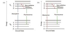
Nian Peng, Kerui Li, Haixia Qiu, Ying Gu, and Defu Chen
SignificancePhotodynamic therapy (PDT) based on photodynamic reaction has been established as a novel treatment modality for cancers and precancerous lesions. PDT adopts the light with an appropriate wavelength in the presence of oxygen to activate a photosensitizer and generate reactive oxygen species (ROS), which then causes localized cell death or tumor necrosis. Concisely, PDT treatment may be described by specifying the administered photosensitizer dose, treatment light dose, and drug-light interval. As a basic component of PDT, the spatial distribution of photosensitizer may directly influence the efficacy of PDT. In vivo quantitative measurement of photosensitizer concentration provides the basis for personalized PDT. In addition to the purpose of treatment, the information on the spatial distribution of a photosensitizer in tissue can be used to identify the tumor tissue and associated margins better.Accurate quantification of in vivo photosensitizer concentration remains challenging. In addition to the photosensitizer, fluorescence also originates from other endogenous fluorophores. Furthermore, intrinsic and instrumental factors also affect the measured fluorescence signal, limiting the ability to make accurate and reliable measurements. Hence, various technologies have been developed to quantify the fluorescence of photosensitizers. This review introduces the instrumentation and intrinsic factors impacting the in vivo quantitative detection of photosensitizer concentrations. Here, recent research progress in the fluorescence correction methods and quantitative detection techniques of photosensitizers are summarized. Finally, the potential challenges and the prospects of quantitative detection techniques of photosensitizer in the clinical translational application of PDT are also briefly discussed.ProgressThe quantitative detection of photosensitizer concentration is a complex process. The measured fluorescence intensity is influenced by instrumental factors, which include excitation light sources, optical components, detectors, and computers (Fig. 4). Moreover, the measured signal is also significantly impacted by tissue optical properties, which include the scattering and absorption of the excitation light and fluorescence emissions (Fig. 5). Consequently, several techniques have been developed to correct the measured fluorescence for endogenous fluorescence, tissue optical properties, and instrumental factors. The correction techniques can be broadly categorized as empirical, Monte Carlo (MC) simulation, and theoretical methods (Fig. 6).The empirical methods have been commonly used to compensate for the attenuation triggered by tissue absorption and scattering on the excitation light and measured fluorescence emissions, which mainly include subtraction and ratio techniques. The empirical methods have the potential to enable near real-time data processing, owing to the inherent simplicity of the proposed methods. MC simulations are most widely used to correct fluorescence measurements, and MC modeling can be used to simulate fluorescence signals collected by an isotropic detector placed on a tissue surface with varying optical properties. The parameters for correction factors can be readily obtained from the MC simulations. The conventional theoretical methods mainly include diffusion theory, modified Beer-Lambert law, and Kubelka-Munk theory. The theoretical methods usually necessitate calculating the transfer function relating intrinsic to measured fluorescence.To date, technologies for fluorescence quantification have used either contact, handheld spectroscopic probes, or non-contact, wide-field imaging systems (Fig. 7). The handheld spectroscopic probes have proven to be an effective technique for precisely quantifying the photosensitizer concentration by utilizing rigorous correction methods (Fig. 8). The handheld fiber-optic probe can quantitatively measure photosensitizer (e.g., chlorine e6) concentration in vivo. Consequently, handheld probes have commonly been used as the “gold standard” for fluorescence qualification. However, it can only measure a small field of view for each acquisition. Wide-field imaging systems allow imaging of the spatial distribution of photosensitizer over a larger area (Fig. 9). This technique provides a map of fast estimation of photosensitizer concentration across the field of view. Despite the aforementioned advantages, fluorescence quantification using a wide-field imaging system remains challenging because fluorescence imaging is highly sensitive to lighting variations and varying distances (e.g., distance between excitation and tissue, distance between tissue and detector).In addition to spectroscopic probes and wide-field systems, several novel quantitative techniques have been proposed for fluorescence quantification, including fluorescence tomography, single-cell resolved microscopic system, portable imaging system, and endoscopic imaging system. Fluorescence tomography enables the 3D spatial distribution information of photosensitizer to be obtained. Single-cell resolved microscopic system is an encouraging technique for imaging tissue at cellular resolution and has the potential to reveal intra-tumor heterogeneity. Portable quantitative fluorescence imaging systems (e.g., smartphone-based systems) provide convenient image collection, computation, and quantitative imaging guidance at the point of care. The endoscopic fluorescence quantitative imaging system is intended for in vivo imaging of internal body organs.Conclusions and ProspectsIn vivo qualification of photosensitizer concentration is crucial for personalized PDT and cancer diagnosis. However, the quantitative detection of photosensitizer concentration is a complex process. Ongoing research attempts to develop the depth-resolved, high-sensitivity, high-resolution optical imaging technique for in vivo real-time quantification of the photosensitizer concentration for pre-, during- and post-PDT.
Feb. 10, 2023Vol. 50 Issue 3 0307201 (2023)
Kang Xu, Tian Zhang, Jinjun Shao, and Xiaochen Dong
SignificancePhotodynamic therapy (PDT) is an effective tumor treatment modality that uses light-activating photosensitizing drugs known as photosensitizers (PSs). PSs selectively accumulate in tumor tissues and convert molecular oxygen in cells and tissues into biotoxic reactive oxygen species (ROS) under specific wavelengths of light irradiation, resulting in oxidative damage to cancer cells. PDT is noninvasive and has low toxicity. It has attracted widespread attention in tumor treatment research. However, there are still challenges in the use of PDT for the treatment of tumors. First, the tissue penetration depth of light is insufficient, which reduces the PDT effect in deep tumor tissue. Second, PDT has poor targeting ability and can easily cause damage to normal tissues. Third, the tumor tissue can scavenge the generated ROS, reducing the therapeutic efficiency of PDT.Compared with normal tissue, tumor tissue has significant pathological changes, showing a characteristic tumor microenvironment, such as weak acidity, hypoxia, protease overexpression, and glutathione (GSH) overexpression. In the design of PSs, overexpressed GSH in the tumor microenvironment is used as a biotarget and trigger factor that can selectively activate the photodynamic activity of PSs accumulated at tumor sites. Therefore, PDT-targeted treatment of tumors can be achieved, and damage to normal tissues can be reduced. However, overexpression of GSH also consumes ROS generated by PS irradiation, which seriously affects the therapeutic efficiency of PSs in tumor phototherapy. In this review, the roles of GSH in organisms are introduced, and GSH-activating and GSH-depleting PSs that have appeared in recent years are described. Finally, the challenges and future development directions of GSH-responsive PSs are discussed.ProgressActivated PSs are only selectively activated in tumors for PDT and have less nonspecific phototoxicity. Under light irradiation, the activable PS maintains quenching in nontumor tissues, produces only a small amount of ROS, and has low tissue toxicity. However, when PSs are activated by specific tumor microenvironmental biological targets, the ROS production capacity can be significantly improved to achieve in situ targeted therapy of tumors. The concentration of GSH at the tumor site is much higher than that in normal cells and extracellular fluid, making GSH an excellent PS trigger. Using GSH as an endogenous stimulator to activate PSs to trigger photodynamic responses at tumor sites selectively can improve the precision of PDT and tumor treatment efficiency and reduce toxic side effects on normal tissues.The design principle of GSH-activated PSs is mainly to regulate the ROS production capacity of the PS through fluorescence resonance energy transfer (FRET) or intramolecular photoelectron transfer (PeT). The disulfide bond (S—S) is broken by the attack of reduced GSH, which can be used as a bonding group to connect the donor and acceptor molecules covalently to achieve intramolecular FRET. The disulfide bond can be used as a linking group to connect the PS molecule with the PDT quencher and realize the selective activation of the PS at high concentrations of GSH in tumor tissue. The introduction of electron donors or acceptors into the molecule to form a PeT system with a chromophore can control the ROS generation capacity of the PSs. 2,4-dinitrobenzenesulfonate (DNBS) is often used as a photodynamic quenching group because of its strong electron absorption ability. The excited state of the PS is quenched by introducing the DNBS group, and the singlet oxygen generation ability is then quenched. Before the PS is activated, it does not exhibit cytotoxicity. After the sulfonyl group in DNBS is cleaved under the induction of GSH, the DNBS group leaves, and the PS is activated, which can generate cytotoxic singlet oxygen. In addition, azo ligands and nitro groups are often used to design GSH-responsive PSs.The high expression of GSH in tumor cells can eliminate exogenous ROS, which is not conducive to the accumulation of exogenous ROS and reduces the efficiency of PDT treatment. Currently, the most common method used to consume GSH is the use of disulfide and highly oxidized metal ions, including manganese (Ⅲ), iron (Ⅲ), and copper (Ⅱ) ions. High-oxidation-state metal ions can consume GSH through redox reactions, and the metal ions themselves are reduced to low-oxidation-state metal ions. The introduction of disulfide bonds and highly oxidized metal ions into PSs can effectively reduce the GSH concentration and improve the therapeutic effect of PDT.Conclusions and ProspectsGSH-activated PSs can be selectively turned on at tumor sites, improving the accuracy of PDT and tumor treatment efficiency and reducing side effects on normal tissues. The consumption of GSH not only solves the problem of ROS scavenging and improves PDT efficacy but also reduces the drug resistance of tumors and improves the therapeutic effect. Although research on GSH-responsive PSs has made significant progress recently, some problems and challenges remain. First, the exact role of GSH in multiple tumors and different tumor stages must be elucidated. Second, GSH deletion leads to the upregulation of related GSH synthetase, potentially negatively affecting tumor therapy. Finally, how to fundamentally inhibit GSH synthesis remains to be studied.
Feb. 10, 2023Vol. 50 Issue 3 0307202 (2023)
Huifang Liu, Dongqin Lei, Fen Qin, Sijia Wang, and Zhenxi Zhang
ObjectiveMelanoma is one of the most immunogenic solid tumors, with strong metastatic characteristics. Although melanoma is curable when detected in a localized form, the long-term survival rates of patients are difficult to improve by any single therapy. The combination of photodynamic therapy (PDT) and immunotherapy has shown encouraging therapeutic efficacy against various cancers due to PDT-induced immunogenic cell death. Natural killer (NK) cell-based immunotherapy has the ability to enhance the anticancer effects of PDT. The NK cell family 2 member D/ligand (NKG2D/NKG2DL) pathway acts as a master switch in the activation of NK cells and mediates the recognition of tumor cells by NK cells. However, tumor cells can produce soluble NKG2DLs via shedding, which mediates the immune escape of tumors. Regulating NKG2D-related molecular pathways can effectively improve the anti-tumor efficacy of NK cells. Further, combined with PDT, NK-related anti-tumor immunity and therapeutic effects could be significantly promoted. In this study, a tumor microenvironment- and light-responsive nanoparticle—MMP-2-responsive polypeptide mixed liposome (MRPL-SC)—was designed to realize synergistic anti-tumor effects of Ce6-mediated PDT and SB-3CT enhanced NKG2D-based immunotherapy, and spatial–temporally controlled release of loaded drugs.MethodsTo synthesize MRPL-SC, DSPE-PEG(3400)-MRP-β-CD with an MMP-2 cleavable sequence was formed via multistep reactions. Then, MRPL-SC, combined with β-cyclodextrin (β-CD) containing SB-3CT to suppress the overexpressed MMP-2 that is tightly associated with the tumor NK-related escape mechanism in the tumor microenvironment and photosensitizer Ce6-loaded liposomes, was prepared using the film dispersion and extrusion method. The multi-responsive release ability of SB-3CT triggered by the MMP-2 abundant tumor microenvironment and chlorin e6 (Ce6) by 660 nm laser irradiation was studied in vitro. Cellular uptake, ROS generation, and the synergistic anti-tumor effects of MRPL-SC-mediated PDT and enhanced NK immunotherapy were studied in A375 cells, followed by the analysis of NKG2DLs expression. In addition, the anti-tumor efficiency and safety of MRPL-SCs were studied in tumor-bearing nude BALB/c mouse models.Results and DiscussionsThe prepared MRPL-SC exhibited a typical liposome structure with a phospholipid bilayer and formed single/double-layer vesicles for drug encapsulating and intracellular delivering with a particle size about 160-170 nm (Fig. 2). Then, the MMP-2-responsive polypeptide linker was hydrolyzed by MMP-2, and SB-3CT encapsulated in β-CD was rapidly released, with accumulation reaching 72.26% after 240 min, indicating the responsiveness of MRPL-SC to the MMP-2 abundant tumor extracellular microenvironment. The released SB-3CT effectively regulated the expression of NKG2DLs and generation of soluble NKG2DLs in tumor tissues by directly inhibiting MMP-2 in situ. Ce6-encapsulated liposomes entered the tumor cells through endocytosis and induced severe cytotoxicity by PDT under 660 nm laser irradiation (Fig.5). In addition, Ce6-PDT also induced NKG2DLs expression, thereby realizing spatiotemporal synergistic enhancement of NK immunity in tumor tissues (Fig. 6). By employing the A375 tumor-bearing nude mice xenograft model, MRPL-SCs showed higher amounts of aggregation in tumor tissue and efficiently suppressed melanoma growth after laser irradiation (Fig. 8) without significant cytotoxic effects in the major organs.ConclusionsIn this study, a novel multi-responsive nanoliposome composed of photosensitizer Ce6-liposomes and MMP-2 antagonist SB-3CT linked by MMP-2 responsive peptide was successfully fabricated via film dispersion and extrusion. The synergistic anti-tumor effect of MRPL-SC-mediated PDT and NK cell immune enhancement was observed in both melanoma cells and nude mouse xenograft models. This study not only developed a new multi-responsive nanocomplex for better integration of PDT and immunotherapy but also elucidated a novel paradigm for NK cell-mediated photodynamic immunotherapy for melanoma.
Feb. 10, 2023Vol. 50 Issue 3 0307203 (2023)
Gege Fu, Yang Lu, Jialin Pan, Xu Li, Chenguang Wang, Xiaomin Liu, and Geyu Lu
ObjectiveTraditional cancer treatments include surgery, radiation therapy, and chemotherapy; however, they have evident side effects. On the contrary, photothermal therapy has attracted considerable attention because of its non-invasive properties, precision, and lower side effects. Photothermal therapy is a treatment method in which a material with high photothermal conversion efficiency is injected into the biological body, and the material converts laser energy into heat energy to kill cancer cells under the irradiation of an external laser source. However, cells in hot environments initiate self-protection measures to resist the damage caused by high temperatures. To optimize the effect of photothermal therapy, a two-dimensional Ti3C2(MXene)-based nano reagent for synergistic photothermal chemotherapy was constructed, quercetin was selected as an anticancer drug, and the inhibitory effect of the nano reagent on tumor cells was explored at the cellular level.MethodsIn this study, LiF/HCl was used to selectively etch the Al layer of Ti3AlC2 to obtain accordion-like multilayer Ti3C2. The obtained wet precipitate was then dissolved in 40 mL of ethanol, sonicated for 80 min to cause initial stratification of the aggregates, washed repeatedly, and thereafter dissolved in deionized water. Finally, the supernatant was obtained as a nanosheet colloidal dispersion by centrifugation and purification after sonication in an ultrasonic cell crusher. Subsequently, the nanosheet surface was modified with the positively charged polymer polypropylene amine hydrochloride to make it positively charged, and PVP-modified quercetin was compounded onto the nanosheet by electrostatic adsorption to form a nanoreagent for synergistic photothermal/chemotherapy treatment. Next, the surface morphology was observed using transmission electron microscopy (TEM), and the composite was analyzed using Zeta potential analysis and absorption spectroscopy. The photothermal effects of the nanosheets and their complexes were subsequently evaluated, and the antitumor effects of this synergistic therapeutic nanoagent were evaluated in cells.Results and DiscussionsThe prepared nanosheets are suitable in size, between about 200-300 nm, and applicable in biomedicine. It can also be observed that quercetin was successfully compounded onto the nanosheets [Fig. 2(c)]. By testing the Zeta potential of the PAH-modified MXene, PVP-modified quercetin, and the final compound, it can be visually confirmed that the final compound has a slightly decreased potential compared to the modified MXene. This indicates that quercetin was successfully complexed to the nanosheet surface [Fig. 2(d)]. Next, the absorption spectra of MXene, quercetin, and MXene@Qu were tested separately, and it was observed that the absorption peak at 261 nm is characteristic to quercetin, and the absorption window of MXene near the NIR appeared in the absorption spectrum of MXene@Qu. This further indicates that the MXene and quercetin were successfully compounded [Fig. 2(e)]. The UV-Vis-NIR absorption spectra revealed the relationship between the absorbance intensity of aqueous Ti3C2 nanosheets and the concentration of the material, which showed the concentration dependence of the absorbance of the composites in the first biological window [Fig. 2(f)]. The extinction coefficient α of the aqueous Ti3C2 nanosheets was calculated to be 24.05 L/(g·cm) according to the Lambert-Beer law, indicating that the prepared Ti3C2 has good near-infrared absorption properties. Subsequently, the concentration and excitation power were changed to observe the concentration- and power-dependent photothermal effects of the nanosheets, indicating that the photothermal effects can be fine-tuned by adjusting the nanosheet concentration or laser intensity. MXene@Qu exhibited good photothermal stability in subsequent heating and cooling cycles. The photothermal conversion efficiency of this nanosheet, obtained by quantitative calculation through one heating-cooling cycle to be 31.34% (Fig. 3). Then, via MTT cytotoxicity assay, it was found that more than 78.91% of the cells of this nanoreagent were killed by laser irradiation, which has a good anti-tumor effect (Fig. 4). In addition, the highest number of dead cells in the MXene@Qu+laser group was qualitatively observed in the live–dead cell staining (Fig. 5).The nanoreagent has excellent anti-tumor effects and achieves good therapeutic results in the in vivo cellular assays, leading to 78.91% cell death. In the future experiments, we will continue to explore the synergistic therapeutic effect of this nanoagent and construct HepG2 tumor-bearing nude mice to explore the effect of photothermal treatment in vivo to achieve the expected effect and effectively improve the efficiency of photothermal treatment.ConclusionsIn this study, we constructed a two-dimensional Ti3C2 (MXene)-based nanoreagent for synergistic photothermal chemotherapy treatment and described the therapeutic effects of the photothermal platform in terms of material properties, in vitro photothermal effects, and intracellular photothermal therapeutic effects. Compared with other photothermal agents, this nanoagent has a high conversion effect (31.34%), good photothermal stability, suitable size, and good hydrophilicity, all of which are in line with the requirements of nanobiotechnology.
Feb. 10, 2023Vol. 50 Issue 3 0307204 (2023)
Yuwei Li, Yu Liu, Lianghua Xie, Kehong Li, Anqi Deng, Wenjie Wu, Qiang Shu, Pengfei Zhao, Weiwei Ke, Rumao Tao, Honghuan Lin, Jianjun Wang, and Feng Jing
Feb. 10, 2023Vol. 50 Issue 3 0316001 (2023)
Chen Chen, Fang Wei, Qingshuai Su, Haoyang Pi, Guofeng Xin, Huimin Wu, Yanguang Sun, Qing Ye, Xiuyou Han, and Haiwen Cai
Feb. 10, 2023Vol. 50 Issue 3 0316002 (2023)
Yihua Hu, Xinyuan Zhang, Fei Han, Yicheng Wang, Shilong Xu, Nanxiang Zhao, Shiyang Chen, Jiaxiang Liu, Jiajie Fang, Xiao Dong, and Ahui Hou
Feb. 10, 2023Vol. 50 Issue 3 0316003 (2023)
© Copyright 2018-2021 | Chinese Laser Press.
All Rights Reserved 沪ICP备15018463号-20