


 View fulltext
Jianying Jing, Kun Liu, Junfeng Jiang, Tianhua Xu, Shuang Wang, and Tiegen Liu
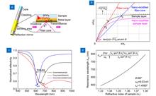
A dispersion model is developed to provide a generic tool for configuring plasmonic resonance spectral characteristics. The customized design of the resonance curve aiming at specific detection requirements can be achieved. According to the model, a probe-type nano-modified fiber optic configurable plasmonic resonance (NMF-CPR) sensor with tip hot spot enhancement is demonstrated for the measurement of the refractive index in the range of 1.3332–1.3432 corresponding to the low-concentration biomarker solution. The new-type sensing structure avoids excessive broadening and redshift of the resonance dip, which provides more possibilities for the surface modification of other functional nanomaterials. The tip hot spots in nanogaps between the Au layer and Au nanostars (AuNSs), the tip electric field enhancement of AuNSs, and the high carrier mobility of the WSe2 layer synergistically and significantly enhance the sensitivity of the sensor. Experimental results show that the sensitivity and the figure of merit of the tip hot spot enhanced fiber NMF-CPR sensor can achieve up to 2995.70 nm/RIU and 25.04 RIU−1, respectively, which are 1.68 times and 1.29 times higher than those of the conventional fiber plasmonic resonance sensor. The results achieve good agreements with numerical simulations, demonstrate a better level compared to similar reported studies, and verify the correctness of the dispersion model. The detection resolution of the sensor reaches up to 2.00×10⁻⁵ RIU, which is obviously higher than that of the conventional side-polished fiber plasmonic resonance sensor. This indicates a high detection accuracy of the sensor. The dense Au layer effectively prevents the intermediate nanomaterials from shedding and chemical degradation, which enables the sensor with high stability. Furthermore, the terminal reflective sensing structure can be used as a practical probe and can allow a more convenient operation.
Jun. 25, 2023Vol. 6 Issue 6 220072 (2023)
Xiaojun Xu, Rui Wang, and Zining Yang
Since the first laser was invented, the pursuit of high-energy lasers (HELs) has always been enthusiastic. The first revolution of HELs was pushed by the fusion of laser and aerospace in the 1960s, with the chemical rocket engines giving fresh impetus to the birth of gas flow and chemical lasers, which finally turned megawatt lasers from dream into reality. Nowadays, the development of HELs has entered the age of electricity as well as the rocket engines. The properties of current electric rocket engines are highly consistent with HELs’ goals, including electrical driving, effective heat dissipation, little medium consumption and extremely light weight and size, which inspired a second fusion of laser and aerospace and motivated the exploration for potential HELs. As an exploratory attempt, a new configuration of diode pumped metastable rare gas laser was demonstrated, with the gain generator resembling an electric rocket-engine for improved power scaling ability.
Jun. 25, 2023Vol. 6 Issue 6 220113 (2023)
Oskar Armbruster, Hannes Pöhl, and Wolfgang Kautek
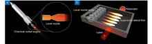
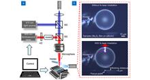
A silver microelectrode with a diameter of 30 µm in an aqueous K2SO4 electrolyte was irradiated with 55 fs and 213 fs laser pulses. This caused the emission of electrons which transiently charged the electrochemical double layer. The two applied pulse durations were significantly shorter than the electron-phonon relaxation time. The laser pulse durations had negligible impact on the emitted charge, which is incompatible with multiphoton emission. On the other hand, the observed dependence of emitted charge on laser fluence and electrode potential supports the thermionic emission mechanism.
Jun. 25, 2023Vol. 6 Issue 6 220170 (2023)
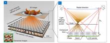
Yan Xing, Xing-Yu Lin, Lin-Bo Zhang, Yun-Peng Xia, Han-Le Zhang, Hong-Yu Cui, Shuang Li, Tong-Yu Wang, Hui Ren, Di Wang, Huan Deng, and Qiong-Hua Wang
Light field 3D display technology is considered a revolutionary technology to address the critical visual fatigue issues in the existing 3D displays. Tabletop light field 3D display provides a brand-new display form that satisfies multi-user shared viewing and collaborative works, and it is poised to become a potential alternative to the traditional wall and portable display forms. However, a large radial viewing angle and correct radial perspective and parallax are still out of reach for most current tabletop light field 3D displays due to the limited amount of spatial information. To address the viewing angle and perspective issues, a novel integral imaging-based tabletop light field 3D display with a simple flat-panel structure is proposed and developed by applying a compound lens array, two spliced 8K liquid crystal display panels, and a light shaping diffuser screen. The compound lens array is designed to be composed of multiple three-piece compound lens units by employing a reverse design scheme, which greatly extends the radial viewing angle in the case of a limited amount of spatial information and balances other important 3D display parameters. The proposed display has a radial viewing angle of 68.7° in a large display size of 43.5 inches, which is larger than the conventional tabletop light field 3D displays. The radial perspective and parallax are correct, and high-resolution 3D images can be reproduced in large radial viewing positions. We envision that this proposed display opens up possibility for redefining the display forms of consumer electronics.
Jun. 25, 2023Vol. 6 Issue 6 220178 (2023)
Zhenyuan Lin, Kuan Liu, Tun Cao, and Minghui Hong
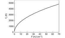
Creation of arbitrary features with high resolution is critically important in the fabrication of nano-optoelectronic devices. Here, sub-50 nm surface structuring is achieved directly on Sb2S3 thin films via microsphere femtosecond laser irradiation in far field. By varying laser fluence and scanning speed, nano-feature sizes can be flexibly tuned. Such small patterns are attributed to the co-effect of microsphere focusing, two-photons absorption, top threshold effect, and high-repetition-rate femtosecond laser-induced incubation effect. The minimum feature size can be reduced down to ~30 nm (λ/26) by manipulating film thickness. The fitting analysis between the ablation width and depth predicts that the feature size can be down to ~15 nm at the film thickness of ~10 nm. A nano-grating is fabricated, which demonstrates desirable beam diffraction performance. This nano-scale resolution would be highly attractive for next-generation laser nano-lithography in far field and in ambient air.
Jun. 25, 2023Vol. 6 Issue 6 230029 (2023)
Perry Ping Shum, Gerd Keiser, Georges Humbert, Dora Juan Juan Hu, A. Ping Zhang, and Lei Su
An ultrasound wave is a kind of acoustic signal with a frequency greater than 20 kHz, which is widely used in diverse fields such as medical imaging diagnosis, nondestructive testing and resource exploration. A variety of ultrasound sensors have been developed for ultrasound detection. Particularly for photoacoustic imaging, specialized ultrasound sensors with high sensitivity, small size, and broad bandwidth are needed. However, achieving such sensor performance still poses a great challenge to the current state-of-the-art in ultrasound sensor technology. A recent work published in Opto-Electronic Advances (DOI: 10.29026/oea.2022.200076) proposes a microfiber-based ultrasound sensor that breaks the limitations of existing ultrasound sensors. Benefiting from the large evanescent field characteristic of microfiber, combined with the coherent detection technology, the proposed sensor realized highly sensitive ultrasound detection and demonstrated excellent performance in high-resolution photoacoustic imaging. The highly sensitive and miniaturized microfiber ultrasound sensor provides a competitive alternative for various applications, such as endoscopic photoacoustic imaging of the intestinal tract and blood vessels in animals.
Jun. 25, 2023Vol. 6 Issue 6 230065 (2023)

Michael John Fanous, and Aydogan Ozcan
A 3D microscopy instrument using flow tomography significantly advances the screening and quantification of intracellular lipid droplets.
Jun. 25, 2023Vol. 6 Issue 6 230083 (2023)
© Copyright 2018-2021 | Chinese Laser Press.
All Rights Reserved 沪ICP备15018463号-20